Accéder au contenu
Merck
Toutes les photos(1)

Key Documents

MABC1132

Sigma-Aldrich

Anti-PD-1 Antibody, clone G4

clone G4, from hamster(Armenian)

Synonyme(s) :

Programmed cell death protein 1, Protein PD-1, mPD-1, CD279

Se connecterpour consulter vos tarifs contractuels et ceux de votre entreprise/organisme


About This Item

Code UNSPSC :
12352203
eCl@ss :
32160702
Nomenclature NACRES :
NA.41

Source biologique

hamster (Armenian)

Forme d'anticorps

purified immunoglobulin

Type de produit anticorps

primary antibodies

Clone

G4, monoclonal

Espèces réactives

mouse

Conditionnement

antibody small pack of 25 μg

Technique(s)

flow cytometry: suitable

Numéro d'accès NCBI

Numéro d'accès UniProt

Modification post-traductionnelle de la cible

unmodified

Informations sur le gène

mouse ... Pdcd1(18566)

Description générale

Protein PD-1, mPD-1, CD279) is encoded by the PDCD1 (also known as PD1) gene (Gene ID: 18566) in murine species. PD-1 is a monomeric inhibitory cell surface receptor involved in the regulation of T-cell function during immunity and tolerance. PD-1 is synthesized with a signal peptide (aa 1-20), which is subsequently cleaved off. The mature form contains an extracellular domain (aa 21-169), a transmembrane domain (aa 170-190), and a cytoplasmic domain (aa 191-288). PD-1 also contains a single N-terminal immunoglobulin variable region (IgV) like domain (aa 31-139). PD-1 and its ligands, PD-L1 and PD-L2 play a key role in the maintenance of peripheral tolerance, a process by which the quiescence of autoreactive mature T cells is maintained. However, tumors and pathogens that cause chronic infections can exploit this pathway to escape T-cell mediated tumor-specific and pathogen-specific immunity. The effector functions of T-cells expressing PD-1 can be downregulated by PD-L1 or PD-L2 expressed by the tumor cells. PD-1 lacks SH2- or SH3-binding motifs on its cytoplasmic tail, but contains the N-terminal sequence VDYGEL that forms an immunoreceptor tyrosine-based inhibition motif (V/I/LxYxxL), which recruits SH2 domain-containing phosphatases. The cytoplasmic tail also contains the C-terminal sequence TEYATI, which forms an immunoreceptor tyrosine-based switch motif (TxYxxL). PD-1 ligation is reported to inhibit the activation of T-cell receptor proximal kinases, which results in attenuation of Lck-mediated phosphorylation of ZAP-70 and initiation of downstream events. PD-1 is also reported to impair the activation of the MEK-ERK MAP kinase pathway by inhibiting activation of PLC- 1 and Ras.

Spécificité

Clone G4 is an Armenian Hamster monoclonal antibody that specifically detects PD-1 in murine cells.

Immunogène

Murine PD-1Ig fusion protein.

Application

Affects Function Analysis: A representative lot detected PD-1 in Affects Funtion applications (Dronca, R.S., et. al. (2016). JCI Insight. 1(6); Hirano, F., et. al. (2005). Cancer Res. 65(3):1089-96; Overacre-Delgoffe, A.E., et. al. (2017). Cell. 169(6):1130; Tsushima, F., et. al. (2006). Blood. 110(1):180-5).

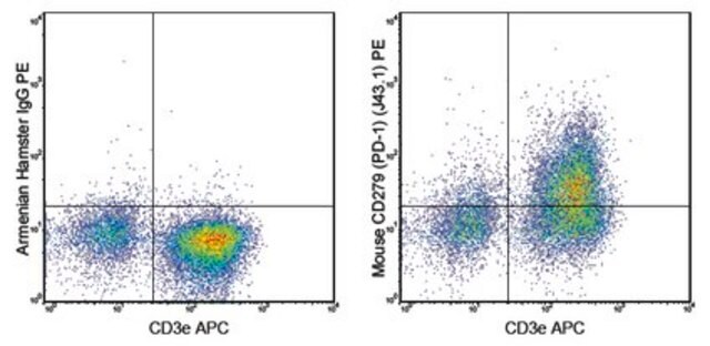
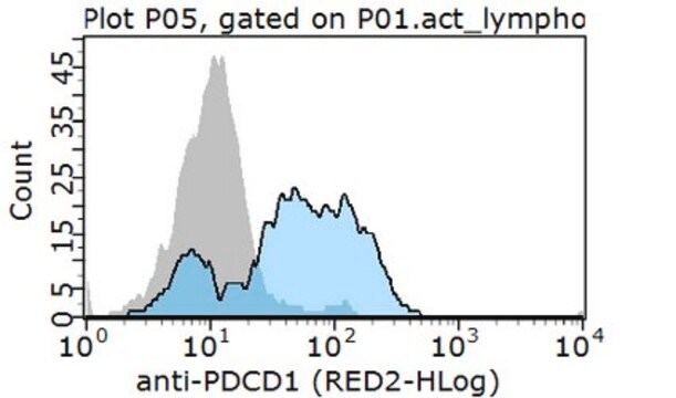
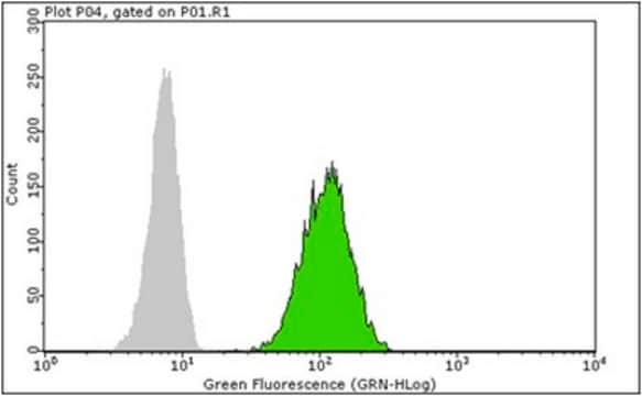
Flow Cytometry Analysis: A representative lot detected PD-1 in Flow Cytometry applications (Hirano, F., et. al. (2005). Cancer Res. 65(3):1089-96).
Detects Programmed cell death protein 1 using this armenian hamster monoclonal Anti-PD-1, clone G4, Cat. No. MABC1132, testted for use in Flow Cytometry and Affects Function.

Qualité

Evaluated by Flow Cytometry in EL4 T lymphoma cells.

Flow Cytometry Analysis: 1 µg of this antibody detected PD-1 in 1X10E6 EL4 T lymphoma cells.

Description de la cible

~31.84 kDa calculated.

Forme physique

Format: Purified

Autres remarques

Concentration: Please refer to lot specific datasheet.

Vous ne trouvez pas le bon produit ?  

Essayez notre Outil de sélection de produits.


Certificats d'analyse (COA)

Recherchez un Certificats d'analyse (COA) en saisissant le numéro de lot du produit. Les numéros de lot figurent sur l'étiquette du produit après les mots "Lot" ou "Batch".

Déjà en possession de ce produit ?

Retrouvez la documentation relative aux produits que vous avez récemment achetés dans la Bibliothèque de documents.

Consulter la Bibliothèque de documents

Ti Wen et al.
Cancer immunology research, 10(2), 162-181 (2021-12-17)
Cytotoxic CD8+ T cells (CTL) are a crucial component of the immune system notable for their ability to eliminate rapidly proliferating malignant cells. However, the T-cell intrinsic factors required for human CTLs to accomplish highly efficient antitumor cytotoxicity are not
Chunhua Chen et al.
JCI insight, 6(8) (2021-04-23)
Although the immune checkpoint role of programmed death ligand 1 (PD-L1) has been established and targeted in cancer immunotherapy, the tumor-intrinsic role of PD-L1 is less appreciated in tumor biology and therapeutics development, partly because of the incomplete mechanistic understanding.

Notre équipe de scientifiques dispose d'une expérience dans tous les secteurs de la recherche, notamment en sciences de la vie, science des matériaux, synthèse chimique, chromatographie, analyse et dans de nombreux autres domaines..

Contacter notre Service technique