Accéder au contenu
Merck
Toutes les photos(1)

Principaux documents

MAB5414

Sigma-Aldrich

Anti-Notch 1 Antibody, extracellular, clone 8G10

culture supernatant, clone 8G10, Chemicon®

Synonyme(s) :

Motch A, mT14, p300

Se connecterpour consulter vos tarifs contractuels et ceux de votre entreprise/organisme


About This Item

Code UNSPSC :
12352203
eCl@ss :
32160702
Nomenclature NACRES :
NA.41

Source biologique

hamster (Syrian)

Niveau de qualité

Forme d'anticorps

culture supernatant

Type de produit anticorps

primary antibodies

Clone

8G10, monoclonal

Espèces réactives

mouse, rat

Fabricant/nom de marque

Chemicon®

Technique(s)

flow cytometry: suitable
immunohistochemistry: suitable
western blot: suitable

Isotype

IgG1

Numéro d'accès NCBI

Numéro d'accès UniProt

Conditions d'expédition

wet ice

Modification post-traductionnelle de la cible

unmodified

Informations sur le gène

human ... NOTCH1(4851)

Description générale

Notch 1 is a type 1 membrane protein that serves as a receptor for the membrane bound ligands jagged-1 and -2 and delta-1 to regulate cell fate determination through the implementation of differentiation, proliferation and apoptotic programs. Upon ligand binding, the Notch intracellular domain (NICD) is released by proteolytic processing and subsequently translocates to the nucleus where it forms a transcriptionally active complex with RBP-J kappa to activate genes of the enhancer of split locus. Notch-1 may also be involved in cell specification and/or differentiation and body segment determination in the postimplantation embryo. In the adult animal, Notch-1 is highly expressed in the brain, lung, and thymus with lower expression levels in the spleen, bone-marrow, spinal cord, eyes, mammary gland, liver, intestine, skeletal muscle, kidney and heart.

Spécificité

Reacts with natural and recombinant Notch1. Recognizes a band migrating at approximately 300 kDa on Western blots of rat and mouse protein extracts. A cross reactive band migrating at approximately 230 kDa is observed in some samples and is believed to correspond to the processed extracellular fragment of Notch 1.

Immunogène

A GST fusion protein comprising amino acids 1299-1492 of mouse Notch 1
Epitope: extracellular

Application

Research Category
Epigenetics & Nuclear Function
Research Sub Category
Transcription Factors
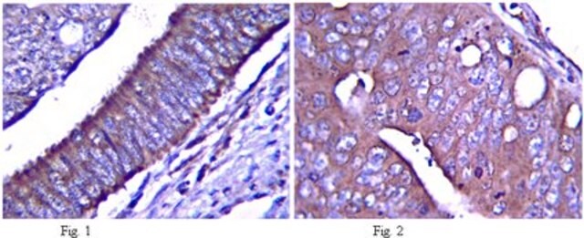
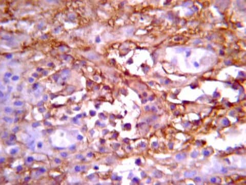
Use Anti-Notch 1 Antibody, extracellular, clone 8G10 (Syrian Hamster Monoclonal Antibody) validated in FC, WB, IHC to detect Notch 1 also known as Motch A, mT14, p300.
Western blot

Immunohistochemistry on paraformaldehyde fixed frozen tissue sections.

Flow Cytometry

Optimal working dilutions must be determined by end user.

Liaison

Replaces: 04-1046

Forme physique

Unpurified tissue culture supernatant from a perfusion system, filtered through a 0.2 micron membrane prior to vialing. Product contains 20% FBS and Ciprofloxacin at final concentration of 10 μg/mL.

Stockage et stabilité

Maintain at -20°C in undiluted aliquots for up to 6 months from date of receipt. Avoid repeated freeze/thaw cycles.

Remarque sur l'analyse

Control
POSITIVE CONTROL: brain, lung, thymus.

Informations légales

CHEMICON is a registered trademark of Merck KGaA, Darmstadt, Germany

Clause de non-responsabilité

Unless otherwise stated in our catalog or other company documentation accompanying the product(s), our products are intended for research use only and are not to be used for any other purpose, which includes but is not limited to, unauthorized commercial uses, in vitro diagnostic uses, ex vivo or in vivo therapeutic uses or any type of consumption or application to humans or animals.

Vous ne trouvez pas le bon produit ?  

Essayez notre Outil de sélection de produits.

Code de la classe de stockage

10 - Combustible liquids


Certificats d'analyse (COA)

Recherchez un Certificats d'analyse (COA) en saisissant le numéro de lot du produit. Les numéros de lot figurent sur l'étiquette du produit après les mots "Lot" ou "Batch".

Déjà en possession de ce produit ?

Retrouvez la documentation relative aux produits que vous avez récemment achetés dans la Bibliothèque de documents.

Consulter la Bibliothèque de documents

Paschalis Kratsios et al.
Circulation research, 106(3), 559-572 (2009-12-17)
The Notch signaling pathway is important for cell-cell communication that controls tissue formation and homeostasis during embryonic and adult life, but the precise cell targets of Notch signaling in the mammalian heart remain poorly defined. To investigate the functional role
Distinct TCR signaling pathways drive proliferation and cytokine production in T cells.
Guy, CS; Vignali, KM; Temirov, J; Bettini, ML; Overacre, AE; Smeltzer, M; Zhang, H; Huppa et al.
Nature Immunology null
Donna Nichol et al.
Blood, 116(26), 6133-6143 (2010-10-16)
Epidermal growth factor-like domain 7 (Egfl7) is important for regulating tubulogenesis in zebrafish, but its role in mammals remains unresolved. We show here that endothelial overexpression of Egfl7 in transgenic mice leads to partial lethality, hemorrhaging, and altered cardiac morphogenesis.
Changhui Ge et al.
Proceedings of the National Academy of Sciences of the United States of America, 105(5), 1539-1544 (2008-01-30)
Mechanisms by which the extracellular domain of Notch1 controls Notch1 signaling are not well defined. Here, we show that the O-fucose glycan in the Notch1 ligand-binding domain regulates the strength of Notch1 signaling during embryogenesis, postweaning growth, and T cell
Sho Morioka et al.
The Journal of cell biology, 204(4), 607-623 (2014-02-19)
TNF activates three distinct intracellular signaling cascades leading to cell survival, caspase-8-mediated apoptosis, or receptor interacting protein kinase 3 (RIPK3)-dependent necrosis, also called necroptosis. Depending on the cellular context, one of these pathways is activated upon TNF challenge. When caspase-8

Notre équipe de scientifiques dispose d'une expérience dans tous les secteurs de la recherche, notamment en sciences de la vie, science des matériaux, synthèse chimique, chromatographie, analyse et dans de nombreux autres domaines..

Contacter notre Service technique