MAB5278
Anti-Tryptophan Hydroxylase/Tyrosine Hydroxylase/Phenylalanine Hydroxylase Antibody
CHEMICON®, mouse monoclonal, PH8
About This Item
Produits recommandés
product name
Anti-Tryptophan Hydroxylase/Tyrosine Hydroxylase/Phenylalanine Hydroxylase Antibody, clone PH8, clone PH8, Chemicon®, from mouse
Source biologique
mouse
Niveau de qualité
Forme d'anticorps
purified immunoglobulin
Type de produit anticorps
primary antibodies
Clone
PH8, monoclonal
Espèces réactives
human
Fabricant/nom de marque
Chemicon®
Technique(s)
immunohistochemistry: suitable
immunoprecipitation (IP): suitable
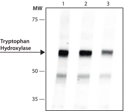
western blot: suitable
Isotype
IgG1
Numéro d'accès NCBI
Conditions d'expédition
wet ice
Modification post-traductionnelle de la cible
unmodified
Informations sur le gène
human ... TPH1(7166)
Description générale
Application
PROTOCOLS
Immunohistochemistry
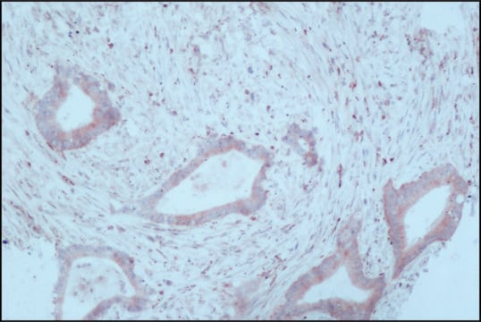
The PH8 Antibody can be used for the immunohistochemical detection of dopaminergic and serotonergic neurons in human and rat brain stem tissue.
1. Tissue should be formalin fixed and stored in formalin prior to use for a minimum of five days.
2. Cryoprotect tissue using 30% sucrose in 0.1M Tris pH 7.4 buffer for 24-72 hours.
3. Cut using a sledge microtome to 50 μm thickness.
4. Wash the tissue samples using Tris buffer prior to commencing the staining procedure.
5. Treat the tissue samples for 3 x 15 minutes in 50% alcohol.
6. Treat the tissue samples for 20 minutes in 50% alcohol and 3% H2O2.
7. Treat tissue for 20 minutes with 10% normal horse serum in Tris buffer. This acts to block endogeneous H2O2 staining.
8. Dilute the PH8 Antibody in Tris buffer, add to the tissue samples and incubate for 1-3 days. Recommended dilutions are 1:2,000-1:10,000. At high antibody concentration all monoaminergic neurons are stained with no distinction between serotonegic and catecholominergic cells. By diluting the antibody concentrate, cell types are distinguishable due to variation in staining intensity. At lower concentrations (1:5,000-1:10,000 dilution) only serotonergic cells will stain.
9. Wash tissue (3 x 15 minutes), then add a biotinylated anti-mouse secondary antibody and incubate on an orbital shaker at room temperature (RT) for 1 hour.
10. Wash tissue (3 x 15 minutes) and incubate on an orbital shaker at RT for 1 hour with the tertiary complex (ELITE KIT, Vector, USA).
11. Wash tissue (3 x 15 minutes) and incubate on an orbital shaker at RT for 10 minutes with Tris buffered diamino-benzidine substrate. Add 0.1% H2O2 and incubate on an orbital shaker at RT for a further 5 minutes.
12. Mount tissue onto gelatinised slides and allow to dry prior to microscopy.
Forme physique
Stockage et stabilité
Informations légales
Vous ne trouvez pas le bon produit ?
Essayez notre Outil de sélection de produits.
Code de la classe de stockage
12 - Non Combustible Liquids
Classe de danger pour l'eau (WGK)
WGK 2
Point d'éclair (°F)
Not applicable
Point d'éclair (°C)
Not applicable
Certificats d'analyse (COA)
Recherchez un Certificats d'analyse (COA) en saisissant le numéro de lot du produit. Les numéros de lot figurent sur l'étiquette du produit après les mots "Lot" ou "Batch".
Déjà en possession de ce produit ?
Retrouvez la documentation relative aux produits que vous avez récemment achetés dans la Bibliothèque de documents.
Notre équipe de scientifiques dispose d'une expérience dans tous les secteurs de la recherche, notamment en sciences de la vie, science des matériaux, synthèse chimique, chromatographie, analyse et dans de nombreux autres domaines..
Contacter notre Service technique


