Accéder au contenu
Merck
Toutes les photos(2)

Key Documents

MAB1913-C

Sigma-Aldrich

Anti-Procollagen Type I Antibody, CT, clone PCIDG10 (Ascites Free)

clone PCIDG10, from mouse

Synonyme(s) :

Collagen alpha-1(I) chain, Procollagen Type I, CT, Alpha-1 type I collagen

Se connecterpour consulter vos tarifs contractuels et ceux de votre entreprise/organisme


About This Item

Code UNSPSC :
12352203
eCl@ss :
32160702
Nomenclature NACRES :
NA.43

Source biologique

mouse

Niveau de qualité

Forme d'anticorps

purified immunoglobulin

Type de produit anticorps

primary antibodies

Clone

PCIDG10, monoclonal

Espèces réactives

mouse, human, rat, guinea pig

Réactivité de l'espèce (prédite par homologie)

bovine (based on 100% sequence homology)

Technique(s)

ELISA: suitable
flow cytometry: suitable
immunocytochemistry: suitable
immunohistochemistry: suitable (paraffin)

Isotype

IgG1κ

Numéro d'accès NCBI

Numéro d'accès UniProt

Conditions d'expédition

dry ice

Modification post-traductionnelle de la cible

unmodified

Informations sur le gène

human ... COL1A1(1277)

Description générale

Collagen alpha-1(I) chain (UniProt P02452; also known as Alpha-1 type I collagen) is encoded by the COL1A1 gene (Gene ID 1277) in human. Collagen is the major component of the extracellular matrix (ECM) and forms the fibrils of tendons, ligaments, and bones. Type I collagen consists of two alpha I chains and one alpha 2 chain. – The mature alpha-1(I) chain is composed mostly of a large triple-helical region (a.a. 179-1192) sandwiched between two nonhelical segments known as the N-terminal telopeptide (a.a. 162-178; numbering based on the prepro-form) and the C-terminal telopeptide (a.a. 1193-1218; numbering based on the prepro-form). Collagen can be extracted from tissue via either enzymatic or non-enzymatic means. Collagen extracted using the proteolytic enzyme pepsin corresponds to the large triple-helical region, referred to as atelocollagen because both the N- and C-terminal telopeptides have been cleaved off by pepsin. On the other hand, collagen preparations obtained with non-enzymatic means (e.g. by acid extraction) have the intact telopeptides at both ends.

Spécificité

Labels carboxy-terminal pro-peptide of collagen type I. Does not stain mature collagen fibers in tissue, but rather is localized intracellularly in cells producing pro-collagen I.

Immunogène

Human pro-collagen I.
Épitope : C-terminal propeptide region

Application

Analyse par immunohistochimie :
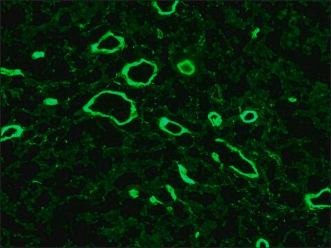
A representative lot detected type I procollagen immunoreactivity in human semitendinosus and gracilis tendon fibroblasts from patients undergoing reconstruction surgery after anterior cruciate ligament (ACL) rupture by fluorescent immunocytochemistry (Bayer, M.L., et al. (2012). Mech Ageing Dev. 133(5):246-254).
Analyse par cytométrie en flux : A representative lot detected PICP+/CD45+ fibrocytes in lung cells from bleomycin-treated mice (Yeager, M.E., et al. (2012).
A representative lot detected higher numbers and percentages of circulating PICP+/CD45+ fibrocytes in peripheral blood samples from children/yound adults with pulmonary hypertension (PH) than in samples from healthy individuals (Reese, C., et al. (2014).
A representative lot detected cytoplasmic type I procollagen immunoreactivity in stromal cells from the lysed functionalis of frozen human menstrual endometria sections (Gaide Chevronnay, H.P., et al. (2009). Endocrinology.
A representative lot detected different age-dependency of procollagen type I C-terminal propeptide (PICP) immunoreactivity in the cruciate ligaments of osteoarthritis-/OA-prone Dunkin-Hartley (DH) guinea pigs and in age-matched Bristol strain 2 (BS2) control guinea pigs (Quasnichka, H.L., et al. (2005). Arthritis Rheum. 52(10):3100-3109).
Domaine de recherche
Structure cellulaire
Sous-domaine de recherche
Molécules d'adhésion cellulaire (CAM)
This Anti-Procollagen Type I Antibody, CT, clone PCIDG10 (Ascites Free) is validated for use in Immunohistochemistry (Paraffin), Immunocytochemistry, Flow Cytometry and ELISA for the detection of Procollagen Type I.

Qualité


A 1:50 dilution of this antibody detected Procollagen Type I in human bone tissue.

Description de la cible

Poids théorique (calculé) : 140 kDa

Forme physique

κ
Format : Produit purifié
Produit purifié sur protéine G

Stockage et stabilité

Stable à -20 °C pendant 1 an à compter de la date de réception.
Recommandations relatives à la manipulation du produit : dès réception, et avant le retrait du bouchon, centrifuger le flacon et mélanger délicatement la solution. Répartir en aliquotes dans des microtubes à centrifuger et conserver ces derniers à -20 °C. Éviter les congélations/décongélations répétées, qui peuvent détériorer les IgG et nuire aux performances du produit.

Autres remarques

Concentration : Voir la fiche technique du lot concerné.

Clause de non-responsabilité

Sauf indication contraire dans notre catalogue ou toute autre documentation associée au(x) produit(s), nos produits sont uniquement destinés à la recherche et ne sauraient être utilisés à d'autres fins, ce qui inclut, sans s'y limiter, les utilisations commerciales non autorisées, les utilisations diagnostiques in vitro, les utilisations thérapeutiques ex vivo ou in vivo, ou tout type de consommation ou d'application chez l'être humain ou chez l'animal.

Vous ne trouvez pas le bon produit ?  

Essayez notre Outil de sélection de produits.

Code de la classe de stockage

12 - Non Combustible Liquids

Classe de danger pour l'eau (WGK)

WGK 1

Point d'éclair (°F)

Not applicable

Point d'éclair (°C)

Not applicable


Certificats d'analyse (COA)

Recherchez un Certificats d'analyse (COA) en saisissant le numéro de lot du produit. Les numéros de lot figurent sur l'étiquette du produit après les mots "Lot" ou "Batch".

Déjà en possession de ce produit ?

Retrouvez la documentation relative aux produits que vous avez récemment achetés dans la Bibliothèque de documents.

Consulter la Bibliothèque de documents

Héloïse P Gaide Chevronnay et al.
Endocrinology, 150(11), 5094-5105 (2009-10-13)
Coupling of focal degradation and renewal of the functional layer of menstrual endometrium is a key event of the female reproductive biology. The precise mechanisms by which the various endometrial cell populations control extracellular matrix (ECM) degradation in the functionalis
J A McDonald et al.
The Journal of clinical investigation, 78(5), 1237-1244 (1986-11-01)
Excessive collagen deposition plays a critical role in the development of fibrosis, and early or active fibrosis may be more susceptible to therapeutic intervention than later stages of scarring. However, at present there is no simple method for assessing the
Yibo Yu et al.
International heart journal, 64(4), 632-640 (2023-07-31)
Atrial fibrillation (AF) is the most common arrhythmia that is harmful to human health. This study aims to explore the relationship between myosin light chain 4 (MYL4) and AF recurrence after radiofrequency ablation (RFA). Patients with AF (n = 85)
Monika L Bayer et al.
Mechanisms of ageing and development, 133(5), 246-254 (2012-03-08)
The aging process of tendon tissue is associated with decreased collagen content and increased risk for injuries. An essential factor in tendon physiology is transforming growth factor-β1 (TGF-β1), which is presumed to be reduced systemically with advanced age. The aim
Helen L Quasnichka et al.
Arthritis and rheumatism, 52(10), 3100-3109 (2005-10-04)
The influence of the cruciate ligaments in spontaneous osteoarthritis (OA) is not understood, although ligament rupture is known to cause secondary OA. Additionally, femoral notch narrowing at the anterior cruciate ligament (ACL) insertion site is associated with disease severity, but

Notre équipe de scientifiques dispose d'une expérience dans tous les secteurs de la recherche, notamment en sciences de la vie, science des matériaux, synthèse chimique, chromatographie, analyse et dans de nombreux autres domaines..

Contacter notre Service technique