ABN991
Anti-phospho GLUT-1 Antibody (Ser226)
from rabbit, purified by affinity chromatography
Synonyme(s) :
Solute carrier family 2, facilitated glucose transporter member 1, Glucose transporter type 1, erythrocyte/brain, GLUT-1, HepG2 glucose transporter, Human T-cell leukemia virus I and II receptor, Receptor for HTLV-1 and HTLV-2, phospho GLUT-1 (Ser226)
About This Item
Produits recommandés
Source biologique
rabbit
Niveau de qualité
Forme d'anticorps
affinity isolated antibody
Type de produit anticorps
primary antibodies
Clone
polyclonal
Produit purifié par
affinity chromatography
Espèces réactives
mouse, human
Réactivité de l'espèce (prédite par homologie)
horse (based on 100% sequence homology), canine (based on 100% sequence homology), bovine (based on 100% sequence homology), Xenopus (based on 100% sequence homology), rat (based on 100% sequence homology), rabbit (based on 100% sequence homology)
Technique(s)
immunocytochemistry: suitable
inhibition assay: suitable (peptide)
western blot: suitable
Numéro d'accès NCBI
Numéro d'accès UniProt
Conditions d'expédition
wet ice
Modification post-traductionnelle de la cible
phosphorylation (pSer226 )
Informations sur le gène
human ... SLC2A1(6513)
Description générale
Immunogène
Application
Western Blotting Analysis: An 1:200 dilution (5 µg/mL) from a representative lot detected TPA-induced phosphorylation of wild-type GLUT-1, but not GLUT-1 with S226A mution in lysates from Rat2 fibroblasts expsssing the respective constructs via retrovirus-mediated transfection (Courtesy of Dr. Richard C. Wang, UT Southwestern Medical Center, Dallas, TX).
Western Blotting Analysis: An 1:200 dilution (5 µg/mL) from a representative lot detected a time-dependent GLUT-1 Ser226 phosphorylation induction in serum-starved human umbilical vein endothelial cells (HUVECs) and human aortic endothelial cells (HAECs) upon VEGF treatment (Courtesy of Dr. Richard C. Wang, UT Southwestern Medical Center, Dallas, TX).
Western Blotting Analysis: A representative lot detected PKC activation-induced GLUT-1 Ser226 phosphorylation in human aortic endothelial cells (HAECs) upon TPA (Cat. No. 500582 & 524400) treatment only the in the absence, but not in the presence, of PKC inhibitor Go 6983 (Cat. No. 365251) (Lee, E.E., et al. (2015). Mol. Cell. 58(5):845-853).
Western Blotting Analysis: A representative lot detected a time-dependent GLUT-1 Ser226 phosphorylation induction in serum-starved human umbilical vein endothelial cells (HUVECs) upon VEGF or angiotensin II treatment (Lee, E.E., et al. (2015). Mol. Cell. 58(5):845-853).
Western Blotting Analysis: A representative lot detected comparable Ser226 phosphorylation induction of exogenously expressed wild-type GLUT-1 or K526E mutant in transfected Rat2 fibroblasts upon PKC activator TPA (Cat. No. 500582 & 524400) treatment (Cat. No. 365251) (Lee, E.E., et al. (2015). Mol. Cell. 58(5):845-853).
Western Blotting Analysis: A representative lot detected PKC activator TPA-induced GLUT-1 Ser226 phosphorylation in serum-starved HeLa, human primary cardiac endothelial cells, EA.hy926 human endothelial cells, and bEnd.3 mouse brain endothelial cells (Lee, E.E., et al. (2015). Mol. Cell. 58(5):845-853).
Western Blotting Analysis: A representative lot detected PKC-catalyzed Ser226 phosphorylation of GST fusion protein containing wild-type GLUT-1 Loop 6 (4th cytoplasmic domain) seuqnce, but not GST-Loop 6 fusions with R223P, R223Q, R223W, or S226A mutation, in in vitro kinase assays (Lee, E.E., et al. (2015). Mol. Cell. 58(5):845-853).
Western Blotting Analysis: A representative lot detected a time-dependent GLUT-1 Ser226 phosphorylation induction in serum-starved human aortic endothelial cells (HAECs) upon VEGF treatment (Lee, E.E., et al. (2015). Mol. Cell. 58(5):845-853).
Immunocytochemistry Analysis: A representative lot detected PKC activation-induced GLUT-1 Ser226 phosphorylation in the membrane ruffles of human umbilical vein endothelial cells (HUVECs) upon TPA (Cat. No. 500582 & 524400) treatment (Lee, E.E., et al. (2015). Mol. Cell. 58(5):845-853).
Note: Process lysate samples by warming at 50°C for 10 minutes prior to gel loading. Avoid heating samples at a temperature higher than 60°C, which can cause aggregation of the target protein.
Qualité
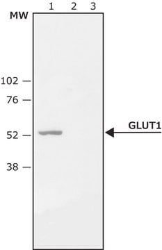
Western Blotting Analysis: 2.0 µg/mL of this antibody detected Ser226 phosphorylated GLUT-1 in lysates from PMA-treated HeLa cells.
Description de la cible
Autres remarques
Vous ne trouvez pas le bon produit ?
Essayez notre Outil de sélection de produits.
Code de la classe de stockage
12 - Non Combustible Liquids
Classe de danger pour l'eau (WGK)
WGK 1
Point d'éclair (°F)
Not applicable
Point d'éclair (°C)
Not applicable
Certificats d'analyse (COA)
Recherchez un Certificats d'analyse (COA) en saisissant le numéro de lot du produit. Les numéros de lot figurent sur l'étiquette du produit après les mots "Lot" ou "Batch".
Déjà en possession de ce produit ?
Retrouvez la documentation relative aux produits que vous avez récemment achetés dans la Bibliothèque de documents.
Notre équipe de scientifiques dispose d'une expérience dans tous les secteurs de la recherche, notamment en sciences de la vie, science des matériaux, synthèse chimique, chromatographie, analyse et dans de nombreux autres domaines..
Contacter notre Service technique


