Accéder au contenu
Merck
Toutes les photos(3)

Key Documents

AB10523

Sigma-Aldrich

Anti-EBF-1 Antibody

from rabbit, purified by affinity chromatography

Synonyme(s) :

Transcription factor COE1, O/E-1, OE-1, Early B-cell factor

Se connecterpour consulter vos tarifs contractuels et ceux de votre entreprise/organisme


About This Item

Code UNSPSC :
12352203
eCl@ss :
32160702
Nomenclature NACRES :
NA.41

Source biologique

rabbit

Niveau de qualité

Forme d'anticorps

affinity isolated antibody

Type de produit anticorps

primary antibodies

Clone

polyclonal

Produit purifié par

affinity chromatography

Espèces réactives

human, mouse

Technique(s)

dot blot: suitable
immunofluorescence: suitable
immunohistochemistry: suitable (paraffin)
western blot: suitable

Numéro d'accès NCBI

Numéro d'accès UniProt

Conditions d'expédition

wet ice

Modification post-traductionnelle de la cible

unmodified

Informations sur le gène

human ... EBF1(1879)

Description générale

The family of Early B-cell Factor (EBF) or Collier/Olf-1/EBF (COE) proteins, EBF1, EBF2, and EBF3, are ubiquitous zinc-binding transcription factors found in many species, including human, Drosophila melanogaster, and Caenorhabditis elegans. EBF1 is abundantly expressed in early B-cells, lymph node, spleen, and adipose tissue. Low levels of EBF1 have also been detected in the brain, heart, skeletal muscle, and kidney. EBF proteins are capable of forming homo- and heterodimers that bind to DNA at specific sites that include the sequence 5′-ATTCCCNNGGGAATT-3′. EBF1, the prototypical member of this family, may employ epigenetic mechanisms, such as DNA methylation and chromatin remodeling, to regulate genes involved in B-cell development such as Pax5. The second member of this family EBF2 is involved in bone development, adipogenesis, and CNS development, processes which may also hinge on EBF1. EBF3 has been characterized as a tumor suppressor protein, due to its role in regulating multiple genes such as those for cyclins and CDKs which control cell growth, differentiation, and apoptosis. However, emerging evidence indicates that all EBF proteins may play a role in the development of various cancers.

Spécificité

This antibody recognizes EBF-1 but not EBF-2 or EBF-3.

Immunogène

KLH-conjugated linear peptide corresponding to mouse EBF-1.

Application

Anti-EBF-1 Antibody is an antibody against EBF-1 for use in Western Blotting, IHC(P), Immunofluorescence, Dot Blot.
Research Category
Neuroscience
Research Sub Category
Developmental Neuroscience
Western Blot Analysis: 2 µg/mL from a representative lot detected EBF-1 in COS-7 cells transfected with EBF-3. (Image courtesy of Dr. Giacomo Consalez, San Raffaele Scientific Institute.)

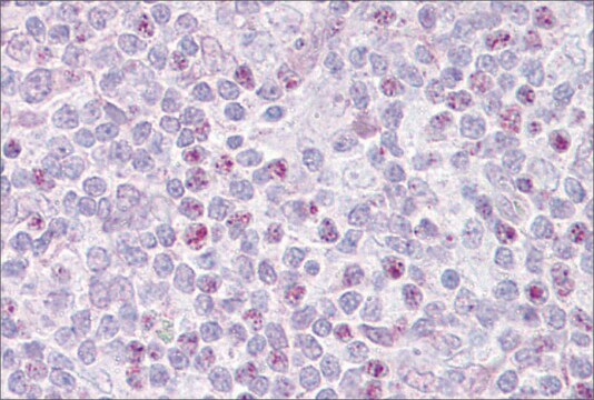
Immunohistochemistry Analysis: A 1:500 dilution from a representative lot detected EBF-1 in mouse cryosections of wild type spinal cord tissue. (Image courtesy of Dr. Giacomo Consalez, San Raffaele Scientific Institute.)

Immunofluorescence Analysis: A 1:500 dilution from a representative lot detected EBF-1 in COS-7 cells transfected with EBF expressing plasmids. (Image courtesy of Dr. Giacomo Consalez, San Raffaele Scientific Institute.)

Dot Blot Analysis: EBF-1, EBF-2, and EBF-3 peptides from a representative lot were probed with Anti-EBF-1 (1:100 dilution). No cross reactivity to peptides for EBF-3 & EBF-2 were observed.

Qualité

Evaluated by Western Blot in Raji cell lysate.

Western Blot Analysis: 2 µg/mL of this antibody detected EBF-1 in 10 µg of Raji cell lysate.

Description de la cible

~66 kDa observed. Uncharacterized bands may appear between ~40 and ~170 kDa in some lysates.

Forme physique

Affinity purified
Purified rabbit polyclonal in buffer containing 0.1 M Tris-Glycine (pH 7.4), 150 mM NaCl with 0.05% sodium azide.

Stockage et stabilité

Stable for 1 year at 2-8°C from date of receipt.

Remarque sur l'analyse

Control
Raji cell lysate

Autres remarques

Concentration: Please refer to the Certificate of Analysis for the lot-specific concentration.

Clause de non-responsabilité

Unless otherwise stated in our catalog or other company documentation accompanying the product(s), our products are intended for research use only and are not to be used for any other purpose, which includes but is not limited to, unauthorized commercial uses, in vitro diagnostic uses, ex vivo or in vivo therapeutic uses or any type of consumption or application to humans or animals.

Vous ne trouvez pas le bon produit ?  

Essayez notre Outil de sélection de produits.

Code de la classe de stockage

12 - Non Combustible Liquids

Classe de danger pour l'eau (WGK)

WGK 1

Point d'éclair (°F)

Not applicable

Point d'éclair (°C)

Not applicable


Certificats d'analyse (COA)

Recherchez un Certificats d'analyse (COA) en saisissant le numéro de lot du produit. Les numéros de lot figurent sur l'étiquette du produit après les mots "Lot" ou "Batch".

Déjà en possession de ce produit ?

Retrouvez la documentation relative aux produits que vous avez récemment achetés dans la Bibliothèque de documents.

Consulter la Bibliothèque de documents

Zhenmeiyu Li et al.
Cell death discovery, 8(1), 301-301 (2022-07-01)
The striatum is the main input structure of the basal ganglia, receiving information from the cortex and the thalamus and consisting of D1- and D2- medium spiny neurons (MSNs). D1-MSNs and D2-MSNs are essential for motor control and cognitive behaviors
Xiaolei Song et al.
Neuroscience bulletin, 37(7), 985-998 (2021-05-21)
Medium spiny neurons (MSNs) in the striatum, which can be divided into D1 and D2 MSNs, originate from the lateral ganglionic eminence (LGE). Previously, we reported that Six3 is a downstream target of Sp8/Sp9 in the transcriptional regulatory cascade of
Andrea Tinterri et al.
The Journal of comparative neurology, 526(3), 397-411 (2017-09-19)
In mammals, thalamic axons are guided internally toward their neocortical target by corridor (Co) neurons that act as axonal guideposts. The existence of Co-like neurons in non-mammalian species, in which thalamic axons do not grow internally, raised the possibility that
Agnieszka Szymula et al.
PLoS pathogens, 14(2), e1006890-e1006890 (2018-02-21)
The Epstein-Barr virus (EBV) nuclear antigen leader protein (EBNA-LP) is the first viral latency-associated protein produced after EBV infection of resting B cells. Its role in B cell transformation is poorly defined, but it has been reported to enhance gene
Chenhe Su et al.
PLoS pathogens, 17(8), e1009834-e1009834 (2021-08-06)
Viruses suppress immune recognition through diverse mechanisms. Epstein-Barr Virus (EBV) establishes latent infection in memory B-lymphocytes and B-cell malignancies where it impacts B-cell immune function. We show here that EBV primary infection of naïve B-cells results in a robust down-regulation

Notre équipe de scientifiques dispose d'une expérience dans tous les secteurs de la recherche, notamment en sciences de la vie, science des matériaux, synthèse chimique, chromatographie, analyse et dans de nombreux autres domaines..

Contacter notre Service technique