SAB4200845
Anti-Fibronectin antibody, Mouse monoclonal
clone IST-3, purified from hybridoma cell culture
Synonym(s):
Mouse Anti-FN
Sign Into View Organizational & Contract Pricing
All Photos(1)
About This Item
UNSPSC Code:
12352203
NACRES:
NA.41
Recommended Products
antibody form
purified from hybridoma cell culture
Quality Level
clone
IST-3
form
liquid
species reactivity
sheep, chicken, human, bovine, goat, dog, rabbit
concentration
~1 mg/mL
technique(s)
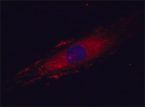
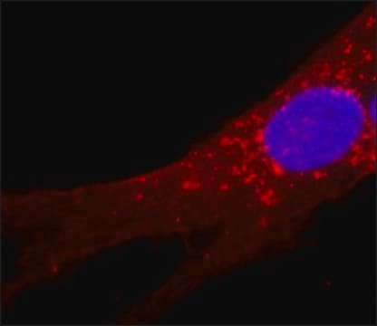
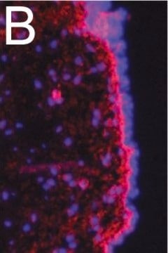
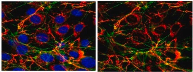
immunofluorescence: 2.5-5 μg/mL using human foreskin fibroblast Hs68 cells
isotype
IgG1
UniProt accession no.
shipped in
dry ice
storage temp.
−20°C
target post-translational modification
unmodified
General description
Two types of Fibronectin are present in vertebrates: soluble plasma Fibronectin and insoluble cellular Fibronectin. The plasma form of Fibronectin (formerly known as cold-insoluble globulin or CIG) is synthesized by hepatocytes, secreted to blood and upon tissue injury, is incorporated into fibrin clots effecting platelet function and mediating hemostasis.8 Cellular Fibronectin is synthesized by many cell types, including fibroblasts, endothelial cells, chondrocytes, synovial cells and myocytes. It is assembled by cells as they migrate into the clot to reconstitute damaged tissue.
Specificity
Monoclonal Anti-Fibronectin antibody specifically recognizes epitope located within the fourth type-three repeat of human plasma fibronectin. The antibody localizes plasma and cellular fibronectin both in its natural and denatured-reduced forms. The product reacts with fibronectin from human, goat, bovine4, porcine5, mouse6, rat7, dog and chicken origin.
Application
The antibody may be used in various immunochemical techniques including Immunoblotting1, ELISA, RIA1-2, Immunofluorescence3 and Immunohistochemistry4.
Biochem/physiol Actions
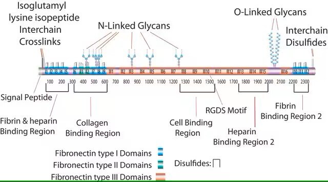
Fibronectin (FN) is a multi-domain glycoprotein composed of two nearly identical disulfide-bound polypeptides with molecular weights of 220-240 KDa. It is a ubiquitous and essential component of the extracellular matrix (ECM) which plays a vital role during tissue repair. Fibronectin functions both as a regulator of cellular processes and as an important scaffolding protein to maintain and direct tissue organization and ECM composition. It is widely expressed by multiple cell types and is critically important in vertebrate development, as demonstrated by the early embryonic lethality of mice with targeted inactivation of the FN gene. Fibronectin is suggested to enhance cell adhesion and spreading and to affect the routes of cell migration both in vivo and in culture.11 It has been shown that upon malignant transformation many cells lose their surface bound fibronectin.
Physical form
Supplied as a solution in 0.01 M phosphate buffered saline pH 7.4, containing 15 mM sodium azide as a preservative.
Storage and Stability
For continuous use, store at 2-8°C for up to one month. For extended storage, freeze in working aliquots. Repeated freezing and thawing is not recommended. If slight turbidity occurs upon prolonged storage, clarify the solution by centrifugation before use. Working dilution samples should be discarded if not used within 12 hours.
Disclaimer
Unless otherwise stated in our catalog our products are intended for research use only and are not to be used for any other purpose, which includes but is not limited to, unauthorized commercial uses, in vitro diagnostic uses, ex vivo or in vivo therapeutic uses or any type of consumption or application to humans or animals.
Storage Class Code
12 - Non Combustible Liquids
WGK
nwg
Flash Point(F)
Not applicable
Flash Point(C)
Not applicable
Certificates of Analysis (COA)
Search for Certificates of Analysis (COA) by entering the products Lot/Batch Number. Lot and Batch Numbers can be found on a product’s label following the words ‘Lot’ or ‘Batch’.
Already Own This Product?
Find documentation for the products that you have recently purchased in the Document Library.
L Zardi et al.
International journal of cancer, 25(3), 325-329 (1980-03-15)
We have produced somatic cell hybrids between mouse plasocytoma cells deficient in hypoxanthine phosphoribosyltransferase (P3 x 63 Ag8) and spleen cells derived from mice immunized with purified human plasma fibronectin. We report herein the properties of monoclonal anti-fibronectin antibodies released
E L George et al.
Development (Cambridge, England), 119(4), 1079-1091 (1993-12-01)
To examine the role of fibronectin in vivo, we have generated mice in which the fibronectin gene is inactivated. Heterozygotes have one half normal levels of plasma fibronectin, yet appear normal. When homozygous, the mutant allele causes early embryonic lethality
H J Simonsz et al.
Documenta ophthalmologica. Advances in ophthalmology, 52(3-4), 409-414 (1982-01-29)
A patient is described with an orbital fistula complicating frontal sinusitis and osteomyelitis of the frontal bone. The fistula was excised, but a fortnight later an acute exacerbation occurred. From the discharging pus a Staphylococcus aureus was cultured and from
Xinglong Zheng et al.
The Journal of biological chemistry, 278(32), 30136-30141 (2003-06-07)
ADAMTS13 consists of a reprolysin-type metalloprotease domain followed by a disintegrin domain, a thrombospondin type 1 motif (TSP1), Cys-rich and spacer domains, seven more TSP1 motifs, and two CUB domains. ADAMTS13 limits platelet accumulation in microvascular thrombi by cleaving the
Yong Mao et al.
Matrix biology : journal of the International Society for Matrix Biology, 24(6), 389-399 (2005-08-03)
The extracellular matrix provides a framework for cell adhesion, supports cell movement, and serves to compartmentalize tissues into functional units. Fibronectin is a core component of many extracellular matrices where it regulates a variety of cell activities through direct interactions
Our team of scientists has experience in all areas of research including Life Science, Material Science, Chemical Synthesis, Chromatography, Analytical and many others.
Contact Technical Service