Skip to Content
Merck
All Photos(2)

Key Documents

16-220

Sigma-Aldrich

Anti-phospho-Ser/Thr-Pro MPM-2 Antibody, Cy5 conjugate

Upstate®, from mouse

Synonym(s):

Anti-MPM-2 Cy5 Antibody, Cy5 Anti-MPM-2, Phospho-Ser/Thr-Pro Detection

Sign Into View Organizational & Contract Pricing


About This Item

UNSPSC Code:
12352203
eCl@ss:
32160702
NACRES:
NA.41

biological source

mouse

Quality Level

conjugate

CY5 conjugate

antibody form

purified antibody

antibody product type

primary antibodies

clone

monoclonal

species reactivity

vertebrates

manufacturer/tradename

Upstate®

technique(s)

immunocytochemistry: suitable
immunofluorescence: suitable
western blot: suitable

isotype

IgG1

shipped in

wet ice

target post-translational modification

phosphorylation (pSer/pThr)

General description

This antibody recognizes a variety of proteins that are phosphorylated during mitosis.
Progression of cells from interphase to mitosis involves alterations in cell structures and activities. The transition from G2 to M phase is induced by M phase-promoting factor, or MPF. In M phase, many proteins are phosphorylated directly by MPF or indirectly by kinases activated by MPF. These M-phase phosphoproteins (MPPs, or MPHOSPHs) permit disassembly of interphase structures and generation of M-phase enzymatic activities and structures. Also, known as Forkhead-related protein FKHL16 or Hepatocyte nuclear factor 3 forkhead homolog 11.

Specificity

Recognizes a phosphorylated epitope (phospho-[Ser/Thr]Pro) found in phosphoproteins such as MAP2, HSP70, cdc25, and DNA topoisomerase IIα, most of which are phosphorylated at the onset of mitosis. The number of phosphoproteins recognized by MPM-2 varies from species to species and with the cell type.

Immunogen

Mitotic human HeLa cell lysate

Application

Anti-phospho-Ser/Thr-Pro Antibody, MPM-2, Cy5 conjugate is a Mouse Monoclonal Antibody for detection of phospho-Ser/Thr-Pro also known as Mitotic protein #2 & has been tested in IF, WB, ICC.
Research Category
Epigenetics & Nuclear Function
Research Sub Category
Cell Cycle, DNA Replication & Repair

Quality

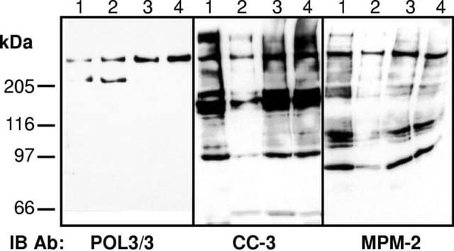
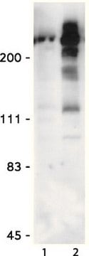
Routinely evaluated by immunoblot on colcemid-treated HeLa cell lysates.

Physical form

100μg Cy5-conjugated mouse IgG1 in 200μl of PBS containing 1% BSA, 0.05% Tween®-20, 0.05% sodium azide.
Protein G Purified

Storage and Stability

1 year at 4°C from date of shipment

Analysis Note

Control
Colcemid-treated HeLa cell lysates

Legal Information

TWEEN is a registered trademark of Croda International PLC
UPSTATE is a registered trademark of Merck KGaA, Darmstadt, Germany

Disclaimer

Unless otherwise stated in our catalog or other company documentation accompanying the product(s), our products are intended for research use only and are not to be used for any other purpose, which includes but is not limited to, unauthorized commercial uses, in vitro diagnostic uses, ex vivo or in vivo therapeutic uses or any type of consumption or application to humans or animals.

Not finding the right product?  

Try our Product Selector Tool.

Storage Class Code

12 - Non Combustible Liquids

WGK

WGK 2

Flash Point(F)

Not applicable

Flash Point(C)

Not applicable


Certificates of Analysis (COA)

Search for Certificates of Analysis (COA) by entering the products Lot/Batch Number. Lot and Batch Numbers can be found on a product’s label following the words ‘Lot’ or ‘Batch’.

Already Own This Product?

Find documentation for the products that you have recently purchased in the Document Library.

Visit the Document Library

Pavlína Víšková et al.
Nature communications, 15(1), 1992-1992 (2024-03-06)
I-Motifs (iM) are non-canonical DNA structures potentially forming in the accessible, single-stranded, cytosine-rich genomic regions with regulatory roles. Chromatin, protein interactions, and intracellular properties seem to govern iM formation at sites with i-motif formation propensity (iMFPS) in human cells, yet
Thomas Blasi et al.
Nature communications, 7, 10256-10256 (2016-01-08)
Imaging flow cytometry combines the high-throughput capabilities of conventional flow cytometry with single-cell imaging. Here we demonstrate label-free prediction of DNA content and quantification of the mitotic cell cycle phases by applying supervised machine learning to morphological features extracted from
Rebecca J Harris et al.
Nature communications, 14(1), 7243-7243 (2023-11-10)
Histone modifications influence the recruitment of reader proteins to chromosomes to regulate events including transcription and cell division. The idea of a histone code, where combinations of modifications specify unique downstream functions, is widely accepted and can be demonstrated in
Xiaowen Sun et al.
Environmental and molecular mutagenesis, 63(5), 230-245 (2022-06-16)
Genotoxicity testing guidelines require the assessment of the clastogenic and aneugenic potential of compounds. While in vitro micronucleus assays detect both types of endpoints, it requires labor-intensive microscopic scoring and does not discriminate between the two modes of actions. Here
Hisayo Nishida-Fukuda et al.
PloS one, 16(4), e0249912-e0249912 (2021-04-15)
HASPIN is a serine/threonine kinase that regulates mitosis by phosphorylating histone H3 at threonine 3. The expression levels of HASPIN in various cancers are associated with tumor malignancy and poor survival, suggesting that HASPIN inhibition may suppress cancer growth. As

Our team of scientists has experience in all areas of research including Life Science, Material Science, Chemical Synthesis, Chromatography, Analytical and many others.

Contact Technical Service