Skip to Content
Merck
All Photos(1)

Key Documents

108R-1

Sigma-Aldrich

CD8 (SP16) Rabbit Monoclonal Antibody

Sign Into View Organizational & Contract Pricing


About This Item

UNSPSC Code:
12352203
NACRES:
NA.41

biological source

rabbit

Quality Level

100
500

conjugate

unconjugated

antibody form

culture supernatant

antibody product type

primary antibodies

clone

SP16, monoclonal

description

For In Vitro Diagnostic Use in Select Regions (See Chart)

form

buffered aqueous solution

species reactivity

human

packaging

vial of 0.1 mL concentrate (108R-14)
vial of 0.5 mL concentrate (108R-15)
bottle of 1.0 mL predilute (108R-17)
vial of 1.0 mL concentrate (108R-16)
bottle of 7.0 mL predilute (108R-18)

manufacturer/tradename

Cell Marque

technique(s)

immunohistochemistry (formalin-fixed, paraffin-embedded sections): 1:100-1:500

isotype

IgG1

control

tonsil

shipped in

wet ice

storage temp.

2-8°C

visualization

membranous

Gene Information

human ... CD8A(925)

General description

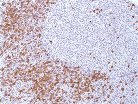
Anti-CD8 is a T-cell marker for the detection of cytotoxic/suppressor lymphocytes. CD8 is also detected on NK cells, most thymocytes, a subpopulation of null cells and bone marrow cells. This antibody, along with other markers, can be used to distinguish between reactive and neoplastic T-cells. Expression of both CD4 (+) and CD8 (+), or lack of expression of both markers in a lymphoid population would be evidence for lymphoma. Although most T-cell lymphoma/leukemias can be either CD8+ or CD8-, CD8 positivity is a factor against Mycosis Fungoides as this entity is negative for CD8. Rarely does anti-CD8 label non-hematolymphoid neoplasms.

Linkage

CD8 Positive Control Slides, Product No. 108S, are available for immunohistochemistry (formalin-fixed, paraffin-embedded sections).

Physical form

Solution in Tris Buffer, pH 7.3-7.7, with 1% BSA and <0.1% Sodium Azide

Preparation Note

Download the IFU specific to your product lot and formatNote: This requires a keycode which can be found on your packaging or product label.

Other Notes

For Technical Service please contact: 800-665-7284 or email: service@cellmarque.com

Legal Information

Cell Marque is a trademark of Merck KGaA, Darmstadt, Germany

Not finding the right product?  

Try our Product Selector Tool.


Choose from one of the most recent versions:

Certificates of Analysis (COA)

Lot/Batch Number

Don't see the Right Version?

If you require a particular version, you can look up a specific certificate by the Lot or Batch number.

Already Own This Product?

Find documentation for the products that you have recently purchased in the Document Library.

Visit the Document Library

Peiguo G Chu et al.
American journal of clinical pathology, 120(1), 64-70 (2003-07-18)
We studied the expression of CD2, CD3, CD4, CD5, CD7, CD8, CD56, and CD138 in 447 cases of common human neoplasms with epithelioid features. CD2, CD3, CD4, and CD8 antigens were detected in none of 447 cases of nonhematopoietic tumors.
Diagnostic Immunohistochemistry: Theranostic and Genomic Applications (2006)
Reactive Lymphadenopathies
Chan J, Tsang W
Pathology of Lymph Nodes, Churchill Livingstone, 81-167 (1996)
G Gaidano et al.
Leukemia, 14(4), 563-566 (2000-04-14)
In recent times, the field of B cell lymphoma histogenesis has progressed rapidly due to the increasing availability of histogenetic markers. Genotypic markers of B cell histogenesis are represented by mutations of IgV and BCL-6 genes, which are somatically acquired
V Bakels et al.
The American journal of pathology, 150(6), 1941-1949 (1997-06-01)
Differentiation between cutaneous pseudo-T-cell lymphomas and cutaneous T-cell lymphomas (CTCLs) may be extremely difficult. In this study, it was investigated whether demonstration of an aberrant phenotype and detection of clonal T-cell receptor gamma (TCR gamma) gene rearrangements can be used

Our team of scientists has experience in all areas of research including Life Science, Material Science, Chemical Synthesis, Chromatography, Analytical and many others.

Contact Technical Service