Skip to Content
Merck
All Photos(1)

Documents

05-173

Sigma-Aldrich

Anti-Calmodulin Antibody

Upstate®, from mouse

Synonym(s):

calmodulin 1, calmodulin 1 (phosphorylase kinase, delta), phosphorylase kinase, delta subunit

Sign Into View Organizational & Contract Pricing


About This Item

UNSPSC Code:
12352203
eCl@ss:
32160702
NACRES:
NA.41

biological source

mouse

Quality Level

antibody form

purified antibody

antibody product type

primary antibodies

clone

monoclonal

species reactivity

pig, mouse, vertebrates, rat, human, bovine

manufacturer/tradename

Upstate®

technique(s)

immunoprecipitation (IP): suitable
western blot: suitable

isotype

IgG1

NCBI accession no.

UniProt accession no.

shipped in

dry ice

target post-translational modification

unmodified

Gene Information

human ... CALM3(808)

General description

Calmodulin (CaM) is a ubiquitous, calcium-binding protein that can bind to and regulate a multitude of different protein targets, thereby affecting many different cellular functions.
CaM mediates processes such as inflammation, metabolism, apoptosis, muscle contraction, intracellular movement, short-term and long-term memory, nerve growth and the immune response. CaM is expressed in many cell types and can have different subcellular locations, including the cytoplasm, within organelles, or associated with the plasma or organelle membranes. Many of the proteins that CaM binds are unable to bind calcium themselves, and as such use CaM as a calcium sensor and signal transducer. CaM undergoes a conformational change upon binding to calcium, which enables it to bind to specific proteins for a specific response. CaM can bind up to four calcium ions, and can undergo post-translational modifications, such as phosphorylation, acetylation, methylation and proteolytic cleavage, each of which can potentially modulate its actions.

Specificity

Also reacts to wheat germ. Wide cross-reactivity expected.
Specific for both Ca2+-bound and Ca2+-free calmodulin, Mr 17 kDa. Does not recognize bovine S100-alpha, S100-beta, troponin C or parvalbumin.

Immunogen

Epitope: a.a. 128-148
Synthetic peptide corresponding to the 21 carboxy-terminal amino acids (128-148) of bovine calmodulin, conjugated with tyroglobulin.

Application

Detect Calmodulin using this Anti-Calmodulin Antibody validated for use in WB & IP.
Immunoprecipitation:
4 μg of a previous lot immunoprecipitated calmodulin from A431 RIPA cell lysate. Radioimmunoassay:
Use 10 μg/mL to detect calmodulin in the range of 200-300 ng.
Research Category
Signaling
Research Sub Category
GPCR, cAMP/cGMP & Calcium Signaling

Quality

Evaluated on human A431 carcinoma cells, RIPA lysates from mouse 3T3 and L6 rat skeletal fibroblasts.

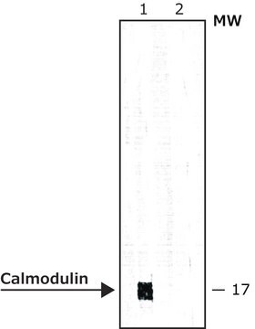
Western Blot Analysis: 0.1-1 μg/mL of this antibody detected calmodulin in RIPA lysates from human A431 carcinoma cells. Previous lots detected calmodulin in RIPA lysates from mouse 3T3 and L6 rat skeletal fibroblasts.

Target description

17 kDa

Linkage

Replaces: 04-1158

Physical form

Format: Purified
Protein G Chromatography
Purified mouse IgG1 in storage buffer containing 0.1 M Tris-glycine, pH 7.4, 0.15 M NaCl, 0.05% sodium azide. Frozen solution.

Storage and Stability

Stable for 1 year at from date of receipt.

Analysis Note

Control
Positive Antigen Control: Catalog #12-301, non-stimulated A431 cell lysate. Add 2.5µL of 2-mercaptoethanol/100µL of lysate and boil for 5 minutes to reduce the preparation. Load 20µg of reduced lysate per lane for minigels.

Other Notes

Concentration: Please refer to the Certificate of Analysis for the lot-specific concentration.

Legal Information

UPSTATE is a registered trademark of Merck KGaA, Darmstadt, Germany

Disclaimer

Unless otherwise stated in our catalog or other company documentation accompanying the product(s), our products are intended for research use only and are not to be used for any other purpose, which includes but is not limited to, unauthorized commercial uses, in vitro diagnostic uses, ex vivo or in vivo therapeutic uses or any type of consumption or application to humans or animals.

Not finding the right product?  

Try our Product Selector Tool.

Storage Class Code

10 - Combustible liquids

WGK

WGK 1


Certificates of Analysis (COA)

Search for Certificates of Analysis (COA) by entering the products Lot/Batch Number. Lot and Batch Numbers can be found on a product’s label following the words ‘Lot’ or ‘Batch’.

Already Own This Product?

Find documentation for the products that you have recently purchased in the Document Library.

Visit the Document Library

Monoclonal antibody to calmodulin: development, characterization, and comparison with polyclonal anti-calmodulin antibodies.
Sacks, D B, et al.
Analytical biochemistry, 194, 369-377 (1991)
Peng Li et al.
International journal of molecular medicine, 37(4), 989-997 (2016-03-05)
The mechanism by which hyperuricemia induced-endothelial dysfunction contributes to cardiovascular diseases (CVDs) is not yet fully understood. In the present study, we used uric acid (UA) to trigger endothelial dysfunction in cultured endothelial cells, and investigated the effects of induced
Human platelets contain estrogen receptor alpha, caveolin-1 and estrogen receptor associated proteins.
Muthuvel Jayachandran, Virginia M Miller, Muthuvel Jayachandran, Virginia M Miller
Platelets null
Induction of TLR4-target genes entails calcium/calmodulin-dependent regulation of chromatin remodeling.
Lai, D; Wan, M; Wu, J; Preston-Hurlburt, P; Kushwaha, R; Grundstrom, T; Imbalzano, AN; Chi, T
Proceedings of the National Academy of Sciences of the USA null
A Lee et al.
Nature, 399(6732), 155-159 (1999-05-21)
Neurotransmitter release at many central synapses is initiated by an influx of calcium ions through P/Q-type calcium channels, which are densely localized in nerve terminals. Because neurotransmitter release is proportional to the fourth power of calcium concentration, regulation of its

Our team of scientists has experience in all areas of research including Life Science, Material Science, Chemical Synthesis, Chromatography, Analytical and many others.

Contact Technical Service