Skip to Content
Merck
All Photos(3)

Documents

WH0004288M1

Sigma-Aldrich

Monoclonal Anti-MKI67 antibody produced in mouse

clone 7B8, purified immunoglobulin, buffered aqueous solution

Synonym(s):

Anti-KIA, Anti-Ki67

Sign Into View Organizational & Contract Pricing


About This Item

MDL number:
UNSPSC Code:
12352203
NACRES:
NA.41

biological source

mouse

conjugate

unconjugated

antibody form

purified immunoglobulin

antibody product type

primary antibodies

clone

7B8, monoclonal

form

buffered aqueous solution

species reactivity

human

technique(s)

immunofluorescence: suitable
indirect ELISA: suitable
western blot: 1-5 μg/mL

isotype

IgG2aκ

GenBank accession no.

UniProt accession no.

shipped in

dry ice

storage temp.

−20°C

target post-translational modification

unmodified

Gene Information

human ... MKI67(4288)

Related Categories

General description

Ki-67 (antigen KI-67) has a gene size of 29,545 bp and it consists of 15 exons and 14 introns. It has a minus strand orientation. This gene is located on human chromosome 10q26.2.
This gene encodes a nuclear protein that is associated with and may be necessary for cellular proliferation. Alternatively spliced transcript variants have been described. A related pseudogene exists on chromosome X. (provided by RefSeq)

Immunogen

MKI67 (NP_002408, 3157 a.a. ~ 3256 a.a) partial recombinant protein with GST tag. MW of the GST tag alone is 26 KDa.

Sequence
RSARQNESSQPKVAEESGGQKSAKVLMQNQKGKGEAGNSDSMCLRSRKTKSQPAASTLESKSVQRVTRSVKRCAENPKKAEDNVCVKKIRTRSHRDSEDI

Application

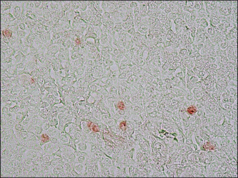
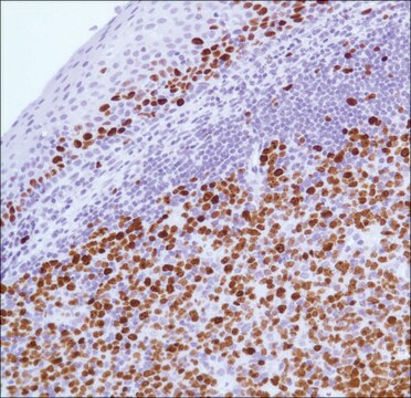
Monoclonal Anti-MKI67 antibody has been used in immunohistochemistry (IHC).

Biochem/physiol Actions

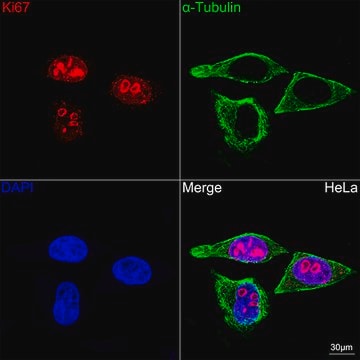
Ki-67 (antigen KI-67) protein acts as a cellular marker for proliferation. This protein plays a major role in the maintenance and regulation of the cell division cycle. Ki-67, a part of the mitotic chromosome periphery, function as a biological surfactant to maintain individual mitotic chromosomes dispersed in the cytoplasm after nuclear envelope disassembly.

Physical form

Solution in phosphate buffered saline, pH 7.4

Legal Information

GenBank is a registered trademark of United States Department of Health and Human Services

Disclaimer

Unless otherwise stated in our catalog or other company documentation accompanying the product(s), our products are intended for research use only and are not to be used for any other purpose, which includes but is not limited to, unauthorized commercial uses, in vitro diagnostic uses, ex vivo or in vivo therapeutic uses or any type of consumption or application to humans or animals.

Not finding the right product?  

Try our Product Selector Tool.

Storage Class Code

10 - Combustible liquids

Flash Point(F)

Not applicable

Flash Point(C)

Not applicable

Personal Protective Equipment

dust mask type N95 (US), Eyeshields, Gloves

Certificates of Analysis (COA)

Search for Certificates of Analysis (COA) by entering the products Lot/Batch Number. Lot and Batch Numbers can be found on a product’s label following the words ‘Lot’ or ‘Batch’.

Already Own This Product?

Find documentation for the products that you have recently purchased in the Document Library.

Visit the Document Library

Customers Also Viewed

Expression of FOXC2, PinX1, Ki-67 and Cyclin D1 in cutaneous cell carcinoma
Zhao H, et al.
Oncology Letters, 14(1), 635-638 (2017)
Prognostic impact of Ki-67 in patients with gastric cancer-the importance of depth of invasion and histologic differentiation
Ko GH, et al.
Medicine, 96(25) (2017)
Ki-67 acts as a biological surfactant to disperse mitotic chromosomes.
Cuylen S, et al.
Nature, 535(7611), 308-308 (2016)
C Schlüter et al.
The Journal of cell biology, 123(3), 513-522 (1993-11-01)
The antigen defined by mAb Ki-67 is a human nuclear protein the expression of which is strictly associated with cell proliferation and which is widely used in routine pathology as a "proliferation marker" to measure the growth fraction of cells
Haiying Zhao et al.
Oncology letters, 14(1), 635-638 (2017-07-12)
We investigated the expression of FOXC2, PinX1, Ki-67 and Cyclin D1 in cutaneous cell carcinoma. We collected 30 cutaneous squamous cell carcinoma (SCC), 30 cutaneous basal cell carcinoma (BCC) and 30 normal skin tissues. The protein expression and gene expression

Our team of scientists has experience in all areas of research including Life Science, Material Science, Chemical Synthesis, Chromatography, Analytical and many others.

Contact Technical Service