Skip to Content
Merck
All Photos(5)

Key Documents

HPA014765

Sigma-Aldrich

Anti-ITPR1 antibody produced in rabbit

Prestige Antibodies® Powered by Atlas Antibodies, affinity isolated antibody, buffered aqueous glycerol solution

Synonym(s):

Anti-ACV, Anti-IP3R1, Anti-Insp3r1, Anti-PPP1R94, Anti-SCA15, Anti-SCA16, Anti-SCA29

Sign Into View Organizational & Contract Pricing


About This Item

UNSPSC Code:
12352203
Human Protein Atlas Number:
NACRES:
NA.41

biological source

rabbit

Quality Level

conjugate

unconjugated

antibody form

affinity isolated antibody

antibody product type

primary antibodies

clone

polyclonal

product line

Prestige Antibodies® Powered by Atlas Antibodies

form

buffered aqueous glycerol solution

species reactivity

human

technique(s)

immunoblotting: 0.04-0.4 μg/mL
immunohistochemistry: 1:500-1:1000

immunogen sequence

KDDFILEVDRLPNETAVPETGESLASEFLFSDVCRVESGENCSSPAPREELVPAEETEQDKEHTCETLLMCIVTVLSHGLRS

UniProt accession no.

shipped in

wet ice

storage temp.

−20°C

target post-translational modification

unmodified

Gene Information

human ... ITPR1(3708)

General description

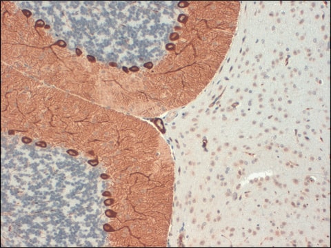
ITPR1 (inositol 1,4,5-trisphosphate receptor, type 1) is a ligand-gated calcium channel, which is present in the endoplasmic reticulum (ER). It either acts as a homotetarmer or forms heterotetramer with type 2 or type 3 inositol 1,4,5-trisphosphate receptors. It is highly expressed in the central nervous system, especially in the Purkinje cells of the cerebellum. This channel contains a coupling/regulatory domain, which consists of phosphorylation sites, a proteolytic cleavage region, ATP-binding sites, and binding sites for multiple proteins. This gene is localized to human chromosome 3p26.

Immunogen

Inositol 1,4,5-trisphosphate receptor type 1 recombinant protein epitope signature tag (PrEST)

Application

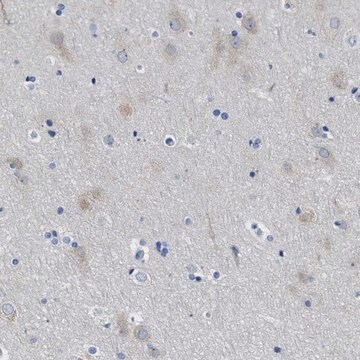
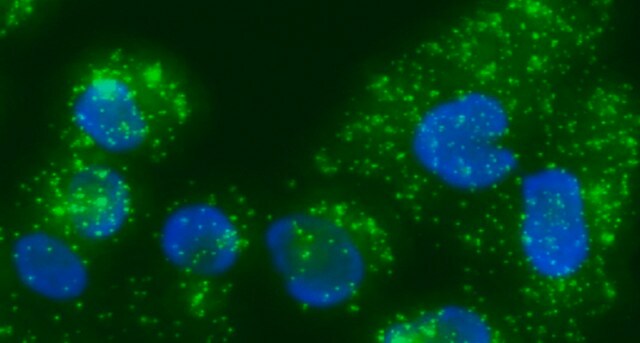
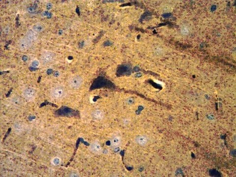
All Prestige Antibodies Powered by Atlas Antibodies are developed and validated by the Human Protein Atlas (HPA) project and as a result, are supported by the most extensive characterization in the industry.

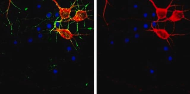
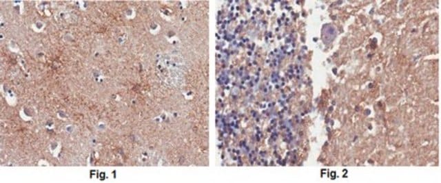
The Human Protein Atlas project can be subdivided into three efforts: Human Tissue Atlas, Cancer Atlas, and Human Cell Atlas. The antibodies that have been generated in support of the Tissue and Cancer Atlas projects have been tested by immunohistochemistry against hundreds of normal and disease tissues and through the recent efforts of the Human Cell Atlas project, many have been characterized by immunofluorescence to map the human proteome not only at the tissue level but now at the subcellular level. These images and the collection of this vast data set can be viewed on the Human Protein Atlas (HPA) site by clicking on the Image Gallery link. We also provide Prestige Antibodies® protocols and other useful information.

Biochem/physiol Actions

ITPR1 (inositol 1,4,5-trisphosphate receptor, type 1) is a Ca2+-channel, which is activated upon binding by inositol 1,4,5-trisphosphate (IP3), and releases Ca2+ from endoplasmic reticulum (ER) into the cytoplasm. This released calcium participates in various Ca2+-dependent signaling and cellular processes. It is activated on death receptor binding or cellular damage, and regulates intracellular calcium levels during apoptosis. Heterozygous deletion in this gene is responsible for the autosomal dominant neurodegenerative disorder spinocerebellar ataxia type 15 (SCA15), which is characterized by late-onset, slow-progressive cerebellar ataxia. It is involved in the activation of T-cells, where it gets phosphorylated at Y353 residue, located at the IP3-binding domain. It plays an essential role in the initial stages of myoblast differentiation, where it induces the expression of myogenin and MEF2 (myocyte enhancer factor 2), which are transcription factors specific to muscles. ITPR1 is also essential for development and maintenance of cerebellum.

Features and Benefits

Prestige Antibodies® are highly characterized and extensively validated antibodies with the added benefit of all available characterization data for each target being accessible via the Human Protein Atlas portal linked just below the product name at the top of this page. The uniqueness and low cross-reactivity of the Prestige Antibodies® to other proteins are due to a thorough selection of antigen regions, affinity purification, and stringent selection. Prestige antigen controls are available for every corresponding Prestige Antibody and can be found in the linkage section.

Every Prestige Antibody is tested in the following ways:
  • IHC tissue array of 44 normal human tissues and 20 of the most common cancer type tissues.
  • Protein array of 364 human recombinant protein fragments.

Linkage

Corresponding Antigen APREST73267

Physical form

Solution in phosphate-buffered saline, pH 7.2, containing 40% glycerol and 0.02% sodium azide

Legal Information

Prestige Antibodies is a registered trademark of Merck KGaA, Darmstadt, Germany

Not finding the right product?  

Try our Product Selector Tool.

Storage Class Code

10 - Combustible liquids

WGK

WGK 1

Flash Point(F)

Not applicable

Flash Point(C)

Not applicable

Personal Protective Equipment

dust mask type N95 (US), Eyeshields, Gloves

Certificates of Analysis (COA)

Search for Certificates of Analysis (COA) by entering the products Lot/Batch Number. Lot and Batch Numbers can be found on a product’s label following the words ‘Lot’ or ‘Batch’.

Already Own This Product?

Find documentation for the products that you have recently purchased in the Document Library.

Visit the Document Library

Lijia Huang et al.
Orphanet journal of rare diseases, 7, 67-67 (2012-09-19)
Congenital nonprogressive spinocerebellar ataxia is characterized by early gross motor delay, hypotonia, gait ataxia, mild dysarthria and dysmetria. The clinical presentation remains fairly stable and may be associated with cerebellar atrophy. To date, only a few families with autosomal dominant
Nikhil deSouza et al.
The Journal of cell biology, 179(5), 923-934 (2007-12-07)
Sustained elevation of intracellular calcium by Ca2+ release-activated Ca2+ channels is required for lymphocyte activation. Sustained Ca2+ entry requires endoplasmic reticulum (ER) Ca2+ depletion and prolonged activation of inositol 1,4,5-trisphosphate receptor (IP(3)R)/Ca2+ release channels. However, a major isoform in lymphocyte
Camia Steinmann et al.
The Journal of biological chemistry, 283(20), 13506-13509 (2008-03-28)
Tumor cells strategically down-regulate Fas receptor expression to evade immune attack and up-regulate expression of Fas ligand to promote apoptosis of infiltrating T lymphocytes. Many pathways leading to apoptotic cell death require calcium release from inositol 1,4,5-trisphosphate receptors (IP3Rs). Here
Fabrice Antigny et al.
Cell calcium, 56(6), 513-521 (2014-12-04)
Cytosolic Ca(2+) signals are fundamental for the early and late steps of myoblast differentiation and are, as in many cells, generated by Ca(2+) release from internal stores as well as by plasma membrane Ca(2+) entry. Our recent studies identified the
Margaret P Adam et al.
GeneReviews(?), 2006 May 30 (Updated 2014 Jun 12) (2014-06-12)
Spinocerebellar ataxia type 15 (SCA15) is characterized by slowly progressive gait and limb ataxia, often in combination with ataxic dysarthria, titubation, upper limb postural tremor, mild hyperreflexia, gaze-evoked nystagmus, and impaired vestibuloocular reflex gain. Onset is between ages seven and

Our team of scientists has experience in all areas of research including Life Science, Material Science, Chemical Synthesis, Chromatography, Analytical and many others.

Contact Technical Service