Skip to Content
Merck
All Photos(5)

Documents

MAB1572

Sigma-Aldrich

Anti-Parvalbumin Antibody

ascites fluid, clone PARV-19, Chemicon®

Synonym(s):

Anti-D22S749

Sign Into View Organizational & Contract Pricing


About This Item

UNSPSC Code:
12352203
eCl@ss:
32160702
NACRES:
NA.41

biological source

mouse

Quality Level

antibody form

ascites fluid

antibody product type

primary antibodies

clone

PARV-19, monoclonal

species reactivity

feline, rabbit, goat, frog, fish, human, pig, bovine, rat, mouse, canine

manufacturer/tradename

Chemicon®

technique(s)

immunocytochemistry: suitable
immunohistochemistry: suitable (paraffin)
western blot: suitable

isotype

IgG1

NCBI accession no.

UniProt accession no.

shipped in

dry ice

target post-translational modification

unmodified

Gene Information

human ... PVALB(5816)

General description

Parvalbumin is a calcium binding albumin protein. It has three EF hand motifs and is structurally related to calmodulin and troponin C. Parvalbumin is localised in fast-contracting muscles, where its levels are highest, and in the brain and some endocrine tissues. Parvalbumin is present in GABAergic interneurons in the nervous system, predominantly expressed by chandelier and basket cells in the cortex.

Specificity

Parvalbumin. The antibody will react with parvalbumin from brain and muscle. By immunoblot it recognizes a protein of 12 kDa. The antibody is directed against an epitope at the first Ca+2-binding site and specifically stains the Ca+2 -bound form of parvalbumin.

Immunogen

Parvalbumin purified from frog muscle

Application

Anti-Parvalbumin Antibody is an antibody against Parvalbumin for use in IC, IH(P) & WB.
Immunohistochemistry:
A previous lot was used by indirect immunoperoxidase at 1:1,000-1:2,000 dilution. The antibody works on formalin fixed, paraffin embedded tissue sections of rat cerebellum from a previous lot. Fixation does not appear to affect MAB1572’s ability to detect parvalbumin.

Immunocytochemistry:
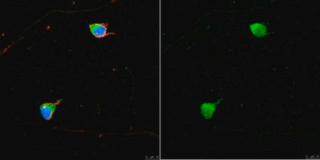
A previous lot of this antibody was used on cultured neurons.

Optimal working dilutions must be determined by the end user.
Research Category
Neuroscience
Research Sub Category
Signaling Neuroscience

Quality

Routinely evaluated by Western Blot on mouse brain lysate.

Western Blot Analysis:
1:1000 dilution of this lot detected Parvalbumin on 10 μg of mouse brain lysate.

Target description

12 kDa

Physical form

Ascites mouse monoclonal IgG1 liquid
Unpurified

Storage and Stability

Stable for 1 year at -20ºC from date of receipt.

Analysis Note

Control
Brain tissue, Cultured neurons

Other Notes

Concentration: Please refer to the Certificate of Analysis for the lot-specific concentration.

Legal Information

CHEMICON is a registered trademark of Merck KGaA, Darmstadt, Germany

Disclaimer

Unless otherwise stated in our catalog or other company documentation accompanying the product(s), our products are intended for research use only and are not to be used for any other purpose, which includes but is not limited to, unauthorized commercial uses, in vitro diagnostic uses, ex vivo or in vivo therapeutic uses or any type of consumption or application to humans or animals.

Not finding the right product?  

Try our Product Selector Tool.

Storage Class Code

12 - Non Combustible Liquids

WGK

nwg

Flash Point(F)

Not applicable

Flash Point(C)

Not applicable


Certificates of Analysis (COA)

Search for Certificates of Analysis (COA) by entering the products Lot/Batch Number. Lot and Batch Numbers can be found on a product’s label following the words ‘Lot’ or ‘Batch’.

Already Own This Product?

Find documentation for the products that you have recently purchased in the Document Library.

Visit the Document Library

Robert Esterberg et al.
The Journal of clinical investigation, 126(9), 3556-3566 (2016-08-09)
Exposure to aminoglycoside antibiotics can lead to the generation of toxic levels of reactive oxygen species (ROS) within mechanosensory hair cells of the inner ear that have been implicated in hearing and balance disorders. Better understanding of the origin of
Stress impairs GABAergic network function in the hippocampus by activating nongenomic glucocorticoid receptors and affecting the integrity of the parvalbumin-expressing neuronal network.
Hu, W; Zhang, M; Czeh, B; Flugge, G; Zhang, W
Neuropsychopharmacology null
Response features of parvalbumin-expressing interneurons suggest precise roles for subtypes of inhibition in visual cortex.
Runyan, CA; Schummers, J; Van Wart, A; Kuhlman, SJ; Wilson, NR; Huang, ZJ; Sur, M
Neuron null
Andrei Avanesov et al.
Methods in cell biology, 100, 153-204 (2010-11-30)
The zebrafish is one of the leading models for the analysis of the vertebrate visual system. A wide assortment of molecular, genetic, and cell biological approaches is available to study zebrafish visual system development and function. As new techniques become
Patricia R Jusuf et al.
The Journal of neuroscience : the official journal of the Society for Neuroscience, 31(7), 2549-2562 (2011-02-18)
Multipotent progenitors in the vertebrate retina often generate clonally related mixtures of excitatory and inhibitory neurons. The postmitotically expressed transcription factor, Ptf1a, is essential for all inhibitory fates in the zebrafish retina, including three types of horizontal and 28 types

Our team of scientists has experience in all areas of research including Life Science, Material Science, Chemical Synthesis, Chromatography, Analytical and many others.

Contact Technical Service