Skip to Content
Merck
All Photos(6)

Documents

05-1003

Sigma-Aldrich

Anti-phospho-Akt (Ser473) Antibody, clone 6F5

clone 6F5, from mouse

Synonym(s):

Protein kinase B, RAC-alpha serine/threonine-protein kinase, murine thymoma viral (v-akt) oncogene homolog-1, rac protein kinase alpha, v-akt murine thymoma viral oncogene homolog 1

Sign Into View Organizational & Contract Pricing


About This Item

UNSPSC Code:
12352203
eCl@ss:
32160702
NACRES:
NA.41

biological source

mouse

Quality Level

antibody form

purified antibody

antibody product type

primary antibodies

clone

6F5, monoclonal

species reactivity

rat, mouse, vertebrates, human

technique(s)

ELISA: suitable
flow cytometry: suitable
immunofluorescence: suitable
immunohistochemistry: suitable
western blot: suitable

isotype

IgG1κ

NCBI accession no.

UniProt accession no.

shipped in

wet ice

target post-translational modification

phosphorylation (pSer473)

Gene Information

human ... AKT1(207)
rat ... Akt1S1(292887)

General description

Akt/PKB is a Ser/Thr kinase and a major known effecter of the PI3 Kinase pathway. It is involved in multiple signaling pathways that relate to many biological processes including glucose metabolism, cell survival/apoptosis, cell cycle control, angiogenesis, differentiation, and cell growth and proliferation. In mammals three isoforms of Akt (Akt1/PKB, Akt2/PKB, and Akt3/PKB)exists. They exhibit a high degree of homology, but differ slightly in the localization of their regulatory phosphorylation sites. Akt is the predominant isoform that is in most tissues and is thought to have a dominant role in growth, survival, embryonic development, and adipocyte differentiation. Akt2 is correlated with the regulation of glucose homeostasis and is the predominant isoform expressed in insulin-responsive tissues. Akt3 is abundant in brain tissue. Each Akt isoform is composed of three functionally distinct regions: an N-terminal Pleckstrin Homology (PH) domain that provides a lipid-binding module to direct Akt to PIP3 at the cell membrane that is necessary for its activation, a central catalytic domain containing Thr308, and a C-terminal hydrophobic motif containing Ser473. The activation of Akt is dependent on a dual regulatory mechanism that requires both its translocation to the plasma membrane and dual phosphorylation on Thr308 and Ser473 by PDK1 and the TORC2 complex, respectively.

Specificity

Expected to react with most species as sequence is 100% homologous with most common vertebrate.
Recognizes Akt only when phosphorylated on Ser473. This antibody does not cross-react with Akt1/PKBα when it is not phosphorylated on Ser473.

Immunogen

Peptide encompassing and including phosphorylated Ser473 of human Akt1.

Application

Anti-phospho-Akt (Ser473) Antibody, clone 6F5 detects level of phospho-Akt (Ser473) & has been published & validated for use in ELISA, WB, FC, IF, IH.
Immunofluroescence Analysis:
EGF-treated A431 cells were fixed, permeabilized, and stained with Anti-phospho-Akt (Ser473), clone 6F5 (2 μg/mL) (red). Cy3-conjugated anti-mouse was used for detection and Phalloidin-AlexaFluor488 (green) was used to stain actin.

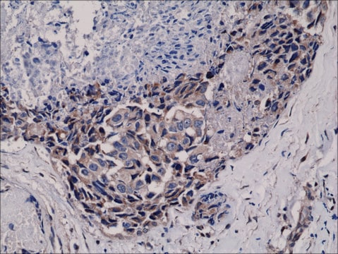
Immunohistochemistry:
Anti-phospho-Akt (Ser473), clone 6F5 was diluted to 1:200, IHC-Select Detection with HRP-DAB.

Flow Cytometry:
Anti-phospho-Akt (Ser473) was used at a 1:800 dilution for this assay.

Quality

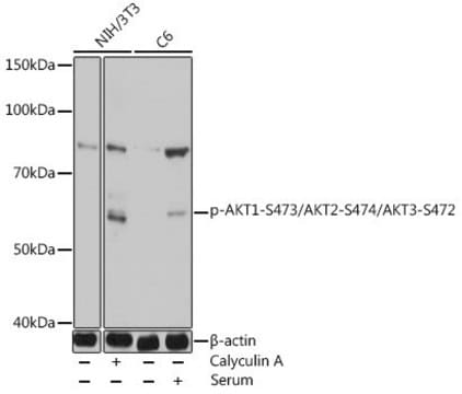
Western Blot Analysis:
0.5-1 μg/mL of this lot detected phosphorylated Akt in lysates from mouse NIH-3T3 fibroblasts treated with 100 ng/mL PDGF for 20 minutes.

Target description

60 kDa

Physical form

Format: Purified
Purified mouse monolconal IgG1κ in 0.1M Tris-Glycine (pH 7.4), 150mM NaCl with 0.05%NaN3

Other Notes

Concentration: Please refer to the Certificate of Analysis for the lot-specific concentration.

Not finding the right product?  

Try our Product Selector Tool.

Storage Class Code

12 - Non Combustible Liquids

WGK

WGK 1

Flash Point(F)

Not applicable

Flash Point(C)

Not applicable


Certificates of Analysis (COA)

Search for Certificates of Analysis (COA) by entering the products Lot/Batch Number. Lot and Batch Numbers can be found on a product’s label following the words ‘Lot’ or ‘Batch’.

Already Own This Product?

Find documentation for the products that you have recently purchased in the Document Library.

Visit the Document Library

Zhicheng Liu et al.
Discover. Oncology, 13(1), 8-8 (2022-02-25)
Colorectal cancer (CRC) is considered to be a leading cause of cancer-related death. Centromere protein O (CENPO) can prevent the separation of sister chromatids and cell death after spindle injury. Nevertheless, the role of CENPO in CRC has not been
Xiaoli Li et al.
The Journal of biological chemistry, 290(5), 2715-2727 (2014-12-17)
Functional maintenance of hematopoietic stem cells (HSCs) is constantly challenged by stresses like DNA damage and oxidative stress. Here we show that the Fanconi anemia protein Fancd2 and stress transcriptional factor Foxo3a cooperate to prevent HSC exhaustion in mice. Deletion
Yuki Miyamoto et al.
Molecular biology of the cell, 26(19), 3489-3503 (2015-08-01)
During early development of the peripheral nervous system, Schwann cell precursors proliferate, migrate, and differentiate into premyelinating Schwann cells. After birth, Schwann cells envelop neuronal axons with myelin sheaths. Although some molecular mechanisms underlying myelination by Schwann cells have been
Claire Marceaux et al.
Journal of cell science, 131(5) (2018-02-09)
ARHGAP19 is a hematopoietic-specific Rho GTPase-activating protein (RhoGAP) that acts through the RhoA/ROCK pathway to critically regulate cell elongation and cytokinesis during lymphocyte mitosis. We report here that, during mitosis progression, ARHGAP19 is sequentially phosphorylated by the RhoA-activated kinases ROCK1
Yanru Wang et al.
BioMed research international, 2021, 6685493-6685493 (2021-03-23)
The vascular injury induced by central venous catheter (CVC) indwelling is the basis for the occurrence and development of CVC-related complications, such as phlebitis, venous thrombosis, and catheter-related infections. Focal adhesion kinase (FAK) and FAK-protein kinase B (AKT) signaling pathway

Articles

Autophagy is a regulated process involved in cell growth, development, and recycling of cytoplasmic components in cells.

Autophagy is a regulated process involved in cell growth, development, and recycling of cytoplasmic components in cells.

Autophagy is a regulated process involved in cell growth, development, and recycling of cytoplasmic components in cells.

Autophagy is a regulated process involved in cell growth, development, and recycling of cytoplasmic components in cells.

Our team of scientists has experience in all areas of research including Life Science, Material Science, Chemical Synthesis, Chromatography, Analytical and many others.

Contact Technical Service