


Based on the information available, this version does recognize the epsilon subunit. This conclusion is drawn from the fact that the CD3 epsilon, also known as the cytoplasmic CD3 antibody, exhibits a cytoplasmic staining pattern with perinuclear Golgi accentuation and very rarely membranous. Typically, this antibody is utilized for identifying T cell and NK cell lymphomas in FFPE tissues (please refer to representative staining photos). While the Cd3 surface antibody is commonly used in flow cytometry for membranous CD3 detection, the CD3 epsilon cytoplasmic is generally used in FFPE tissue, as is often done for IHC.
103R-9
CD3 (MRQ-39) Rabbit Monoclonal Antibody
Se connecterpour consulter vos tarifs contractuels et ceux de votre entreprise/organisme
Sélectionner une taille de conditionnement
1 ML
172,00 €
0.1 ML
189,00 €
7 ML
352,00 €
0.5 ML
465,00 €
1 ML
696,00 €
Sélectionner une taille de conditionnement
Changer de vue
1 ML
172,00 €
0.1 ML
189,00 €
7 ML
352,00 €
0.5 ML
465,00 €
1 ML
696,00 €
About This Item
Code UNSPSC :
12352203
Nomenclature NACRES :
NA.41
Produits recommandés
Source biologique
rabbit
Niveau de qualité
100
500
Conjugué
unconjugated
Forme d'anticorps
culture supernatant
Type de produit anticorps
primary antibodies
Clone
MRQ-39, monoclonal
Description
For In Vitro Diagnostic Use in Select Regions (See Chart)
Forme
buffered aqueous solution
Espèces réactives
human
Conditionnement
pkg of 0.1 mL concentrate (103r-94)
pkg of 0.5 mL concentrate (103R-95)
pkg of 1.0 mL concentrate (103R-96)
pkg of 1.0 mL predilute (103R-97)
pkg of 7.0 mL predilute (103R-98)
Fabricant/nom de marque
Cell Marque®
IVD
for in vitro diagnostic use
Technique(s)
immunohistochemistry (formalin-fixed, paraffin-embedded sections): 1:100-1:500 (concentrated)
Isotype
IgG1
Contrôle
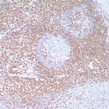
tonsil
Conditions d'expédition
wet ice
Température de stockage
2-8°C
Visualisation
membranous
Informations sur le gène
human ... CD3E(916)
Catégories apparentées
Description générale
Anti-CD3 has been considered the best all around T-cell marker. This antibody reacts with an antigen present in early thymocytes. The positive staining of this marker may represent a sign of early commitment to the T-Cell lineage.
Qualité
 IVD |  IVD |  IVD |  RUO |
Liaison
CD3 Positive Control Slides, Product No. 103S, are available for immunohistochemistry (formalin-fixed, paraffin-embedded sections).
Forme physique
Solution in Tris Buffer, pH 7.3-7.7, with 1% BSA and <0.1% Sodium Azide
Notes préparatoires
Download the IFU specific to your product lot and formatNote: This requires a keycode which can be found on your packaging or product label.
Autres remarques
For Technical Service please contact: 800-665-7284 or email: [email protected]
Informations légales
Cell Marque is a registered trademark of Merck KGaA, Darmstadt, Germany
Vous ne trouvez pas le bon produit ?
Essayez notre Outil de sélection de produits.
Faites votre choix parmi les versions les plus récentes :
Certificats d'analyse (COA)
Lot/Batch Number
Vous ne trouvez pas la bonne version ?
Si vous avez besoin d'une version particulière, vous pouvez rechercher un certificat spécifique par le numéro de lot.
Déjà en possession de ce produit ?
Retrouvez la documentation relative aux produits que vous avez récemment achetés dans la Bibliothèque de documents.
SM Denning, et al.
Leucocyte Typing III, 144-147 (1987)
E A Clark et al.
Immunology today, 10(7), 225-228 (1989-07-01)
During 1987, striking advances were made in defining the receptors and ligands for cell-to-cell adhesion interactions involving leukocytes. In 1988, two major leukocyte differentiation antigens, CD10 (cALLA) and CD45 (LCA, T200), were shown to be enzymes while two other markers
P C Beverley et al.
European journal of immunology, 11(4), 329-334 (1981-04-01)
The properties of human lymphocyte fractions isolated either by sheep red cell(E) rosetting or by fluorescence-activated cell sorting after staining with UCHT1 monoclonal anti-T cell antibody have been compared. Two populations of E+ cells with very different phenotype and function
D Campana et al.
Journal of immunology (Baltimore, Md. : 1950), 138(2), 648-655 (1987-01-15)
Anti-CD3 (T3) Ab reacting with different proportions of thymocytes (anti-CD3a: UCHT1, anti-CD3b: T10B9, and anti-CD3c: OKT3) were tested for cytoplasmic (cCD3) and membrane (mCD3) expression in the bone marrow, thymus, and blood in man and selected primates. The expression of
Kennosuke Karube et al.
The American journal of surgical pathology, 27(10), 1366-1374 (2003-09-26)
We studied the morphologic, immunohistochemical, and clinical characteristics of 158 cases of lymphoblastic lymphoma. Based on immunophenotyping and cell lineage, cases were classified into B-cell type (CD20,CD19 or CD79a+, n = 53), T-cell type (surface CD3+, n = 84), and
-
Could you confirm if the CD3 clone (Catalog #103R-96) recognizes epsilon?
1 réponse-
Utile ?
-
Filtres actifs
Notre équipe de scientifiques dispose d'une expérience dans tous les secteurs de la recherche, notamment en sciences de la vie, science des matériaux, synthèse chimique, chromatographie, analyse et dans de nombreux autres domaines..
Contacter notre Service technique