Accéder au contenu
Merck
Toutes les photos(3)

Documents

MABN24

Sigma-Aldrich

Anti-pan-Shank Antibody, clone N23B/49

clone N23B/49, from mouse

Synonyme(s) :

SH3 and multiple ankyrin repeat domains 2, SH3 and multiple ankyrin repeat domains protein 2, proline-rich synapse associated protein 1, cortactin SH3 domain-binding protein, Cortactin-binding protein 1, Proline-rich synapse-associated protein 1, cortact

Se connecterpour consulter vos tarifs contractuels et ceux de votre entreprise/organisme


About This Item

Code UNSPSC :
12352203
eCl@ss :
32160702
Nomenclature NACRES :
NA.41

Source biologique

mouse

Niveau de qualité

Forme d'anticorps

purified antibody

Type de produit anticorps

primary antibodies

Clone

N23B/49, monoclonal

Espèces réactives

mouse, human, rat

Technique(s)

immunohistochemistry: suitable (paraffin)
western blot: suitable

Isotype

IgG1κ

Numéro d'accès NCBI

Numéro d'accès UniProt

Conditions d'expédition

wet ice

Modification post-traductionnelle de la cible

unmodified

Informations sur le gène

human ... SHANK2(22941)

Description générale

Shank1, Shank2, and Shank3 make up a family of proteins that may function as molecular scaffolds in the postsynaptic density (PSD). Shank contains multiple domains for protein-protein interaction including ankyrin repeats, an SH3 domain, a PSD-95/Dlg/ZO-1 domain, a sterile α motif domain, and a proline-rich region. Alternative splicing in the Shank family may be a mechanism that regulates the molecular structure of Shank and the spectrum of Shank-interacting proteins in the PSDs of adult and developing brain.

Immunogène

Recombinant protein consisting of SH3/PDZ domain of rat Shank2.

Application

Detect pan-Shank using this Anti-pan-Shank Antibody, clone N23B/49 validated for use in WB, IH(P).
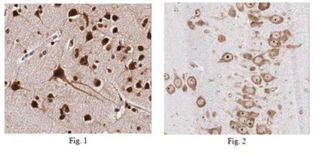
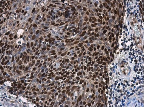
Immunohistochemistry Analysis: 1:500 dilution from a previous lot detected Shank in rat cerebellum tissue.

Western Blot Analysis: A previous lot of this antibody detected Shank in extracts of COS-1 cells transiently transfected with Shank1, Shank2 or Shank3 plasmids. Courtesy of James Trimmer, UC Davis/NIH NeuroMab Facility.
Research Category
Neuroscience
Research Sub Category
Synapse & Synaptic Biology

Qualité

Evaluated by Western Blot in rat brain membrane tissue lysate.

Western Blot Analysis: 2 µg/mL of this antibody detected Shank on 10 µg of rat brain membrane tissue lysate.

Description de la cible

~160 kDa observed. Other isoforms may be observed in some lysates.

Forme physique

Format: Purified
Protein G
Purified mouse monoclonal IgG1κ in buffer containing 0.1 M Tris-Glycine (pH 7.4), 150 mM NaCl with 0.05% sodium azide.

Stockage et stabilité

Stable for 1 year at 2-8°C from date of receipt.

Remarque sur l'analyse

Control
Rat brain membrane tissue lysate

Autres remarques

Concentration: Please refer to the Certificate of Analysis for the lot-specific concentration.

Clause de non-responsabilité

Unless otherwise stated in our catalog or other company documentation accompanying the product(s), our products are intended for research use only and are not to be used for any other purpose, which includes but is not limited to, unauthorized commercial uses, in vitro diagnostic uses, ex vivo or in vivo therapeutic uses or any type of consumption or application to humans or animals.

Not finding the right product?  

Try our Outil de sélection de produits.

Code de la classe de stockage

12 - Non Combustible Liquids

Classe de danger pour l'eau (WGK)

WGK 1

Point d'éclair (°F)

Not applicable

Point d'éclair (°C)

Not applicable


Certificats d'analyse (COA)

Recherchez un Certificats d'analyse (COA) en saisissant le numéro de lot du produit. Les numéros de lot figurent sur l'étiquette du produit après les mots "Lot" ou "Batch".

Déjà en possession de ce produit ?

Retrouvez la documentation relative aux produits que vous avez récemment achetés dans la Bibliothèque de documents.

Consulter la Bibliothèque de documents

Marián Haburčák et al.
Frontiers in synaptic neuroscience, 14, 995474-995474 (2022-10-18)
The Spontaneously Hypertensive Rat (SHR) has increased sympathetic drive to the periphery that precedes and contributes to the development of high blood pressure, making it a useful model for the study of neurogenic hypertension. Comparisons to the normotensive Wistar Kyoto
Kenneth R Myers et al.
Frontiers in molecular neuroscience, 15, 1020949-1020949 (2022-10-18)
Dendritic spines are small actin-rich protrusions essential for the formation of functional circuits in the mammalian brain. During development, spines begin as dynamic filopodia-like protrusions that are then replaced by relatively stable spines containing an expanded head. Remodeling of the
Yusuke Hatanaka et al.
Scientific reports, 5, 16102-16102 (2015-11-05)
Late-onset neurodegenerative diseases are characterized by neurological symptoms and progressive neuronal death. Accumulating evidence suggests that neuronal dysfunction, rather than neuronal death, causes the symptoms of neurodegenerative diseases. However, the mechanisms underlying the dysfunction that occurs prior to cell death

Notre équipe de scientifiques dispose d'une expérience dans tous les secteurs de la recherche, notamment en sciences de la vie, science des matériaux, synthèse chimique, chromatographie, analyse et dans de nombreux autres domaines..

Contacter notre Service technique