Skip to Content
Merck
All Photos(1)

Documents

04-1573

Sigma-Aldrich

Anti-Ataxin-7 Antibody, clone 3SCA-1C1

ascites fluid, clone 3SCA-1C1, from mouse

Synonym(s):

ADCAII, OPCA3, SCA7

Sign Into View Organizational & Contract Pricing


About This Item

UNSPSC Code:
12352203
eCl@ss:
32160702
NACRES:
NA.41

biological source

mouse

Quality Level

antibody form

ascites fluid

antibody product type

primary antibodies

clone

3SCA-1C1, monoclonal

species reactivity

rat, mouse

species reactivity (predicted by homology)

human (immunogen homology)

technique(s)

western blot: suitable

isotype

IgG1κ

NCBI accession no.

UniProt accession no.

shipped in

dry ice

target post-translational modification

unmodified

Gene Information

human ... ATXN7(6314)

General description

Ataxin-7 is a protein of unknown function. However, since this protein contains a nuclear localization sequence and is found to be localized in the nucleus, it has been postulated to be a potential transcription factor. Spinocerebellar ataxia-7 (one of a group of hereditary neurodegenrative diseases) is caused by an expanded CAG repeat in the gene encoding ataxin-7 (the diseased allele associated with Spinocerebellar ataxia-7, contains 38-130 CAG repeats (near the N-terminus), compared to 7-17 in the normal allele). In cells where there is a mutation of the SCA7 gene, ataxin-7 accumulates in intranuclear inclusions and can result in cell death. Spinocerebellar ataxia (SCA) is one of a group of genetic disorders characterized by slowly progressive incoordination of gait and often associated with poor coordination of hands, speech, and eye movements. Frequently, atrophy of the cerebellum occurs.

Specificity

This antibody recognizes Ataxin-7 protein.

Immunogen

Epitope: Epitope mapped between aa 66 and aa 119.
Recombinant human Ataxin 7 as a fusion protein (aa 1 to aa 229).

Application

Anti-Ataxin-7 Antibody, clone 3SCA-1C1 is an antibody against Ataxin-7 for use in WB.
ELISA: A 1:500-1:5,000 dilution of a previous lot of 04-1573 was used in ELISA, as reported by an outside laboratory. Immunocytochemistry: A 1:500-1:5,000 dilution of a previous lot of 04-1573 was used in IC, as reported by an outside laboratory.
Research Category
Neuroscience

Neuroscience
Research Sub Category
Signaling Neuroscience

Neurodegenerative Diseases

Quality

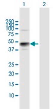
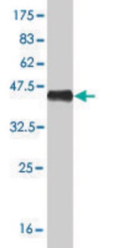
Evaluated by Western Blotting of Rat brain lysate. Western Blotting: Representative lot data: A 1:1000 dilution of this antibody detected Ataxin-7 in 10ug of Rat brain tissue lysate.

Target description

96 kDa Calculated

Physical form

Unpurified
Unpurified mouse IgG1, k antibody in ascites fluid without preservatives.

Storage and Stability

Stable for 1 year at -20°C from date of receipt.
Handling Recommendations: Upon receipt and prior to removing the cap, centrifuge the vial and gently mix the solution. Aliquot into microcentrifuge tubes and store at -20°C. Avoid repeated freeze/thaw cycles, which may damage IgG and affect product performance.

Analysis Note

Control
Rat brain tissue lysate.

Disclaimer

Unless otherwise stated in our catalog or other company documentation accompanying the product(s), our products are intended for research use only and are not to be used for any other purpose, which includes but is not limited to, unauthorized commercial uses, in vitro diagnostic uses, ex vivo or in vivo therapeutic uses or any type of consumption or application to humans or animals.

Not finding the right product?  

Try our Product Selector Tool.

Storage Class Code

12 - Non Combustible Liquids

WGK

nwg

Flash Point(F)

Not applicable

Flash Point(C)

Not applicable


Certificates of Analysis (COA)

Search for Certificates of Analysis (COA) by entering the products Lot/Batch Number. Lot and Batch Numbers can be found on a product’s label following the words ‘Lot’ or ‘Batch’.

Already Own This Product?

Find documentation for the products that you have recently purchased in the Document Library.

Visit the Document Library

S Vijayalingam et al.
Journal of virology, 90(2), 1070-1079 (2015-11-13)
The cell-transforming activity of human adenovirus 5 (hAd5) E1A is mediated by the N-terminal half of E1A, which interacts with three different major cellular protein complexes, p300/CBP, TRRAP/p400, and pRb family members. Among these protein interactions, the interaction of pRb

Our team of scientists has experience in all areas of research including Life Science, Material Science, Chemical Synthesis, Chromatography, Analytical and many others.

Contact Technical Service