Skip to Content
Merck
All Photos(3)

Key Documents

SAB4200341

Sigma-Aldrich

Anti-Golph3 (N-terminal) antibody produced in rabbit

~1.0 mg/mL, affinity isolated antibody

Synonym(s):

Anti-GOPP1, Anti-GPP34, Anti-Golgi peripheral membrane protein 1, 34 kDa, Anti-Golgi phosphoprotein 3 (coat-protein), Anti-Golgi-associated protein; Mitochondrial DNA absence factor, Anti-MIDAS

Sign Into View Organizational & Contract Pricing


About This Item

UNSPSC Code:
12352203
NACRES:
NA.41

biological source

rabbit

Quality Level

conjugate

unconjugated

antibody form

affinity isolated antibody

antibody product type

primary antibodies

clone

polyclonal

form

buffered aqueous solution

mol wt

antigen ~34 kDa

species reactivity

mouse, human

concentration

~1.0 mg/mL

technique(s)

immunoprecipitation (IP): 5-10 μg using lysates of human HeLa cells
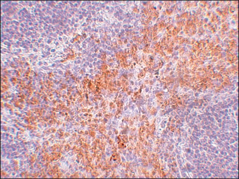
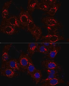
indirect immunofluorescence: 2.5-5.0 μg/mL using mouse 3T3 cells
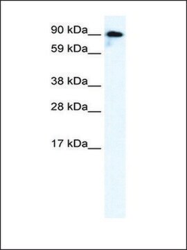
western blot: 1-2 μg/mL using whole extracts of human HeLa cells

UniProt accession no.

shipped in

dry ice

storage temp.

−20°C

target post-translational modification

unmodified

Gene Information

human ... GOLPH3(64083)
mouse ... Golph3(66629)
rat ... Golph3(78961)

General description

Golph3 (Golgi phosphoprotein 3), also known as coat protein GPP34, is a peripheral membrane protein of the Golgi stack, that localizes to the trans-Golgi network. It is an oncogene that is commonly targeted for amplification in human cancer and in cancer cell lines.

Immunogen

peptide corresponding to the N-terminal region of human Golph3, conjugated to KLH. The corresponding sequence is identical in mouse, rat, monkey and bovine.

Application

Anti-Golph3 (N-terminal) antibody produced in rabbit has been used in:
  • immunoprecipitation
  • immunofluorescence
  • immunoblotting

Biochem/physiol Actions

Golph3 (Golgi phosphoprotein 3) is important for Golgi trafficking and morphology by interacting with the unconventional myosin MYO18A, linking Golgi membranes to the actin cytoskeleton. It is a phosphatidylinositol-4-phosphate (PtdIns(4)P binding protein required for Golgi function. GOLPH3 overexpression is correlated with hyperactivation of mTOR signaling, in human cells.

Physical form

Solution in 0.01 M phos­phate buffered saline, pH 7.4, containing 15 mM sodium azide.

Disclaimer

Unless otherwise stated in our catalog or other company documentation accompanying the product(s), our products are intended for research use only and are not to be used for any other purpose, which includes but is not limited to, unauthorized commercial uses, in vitro diagnostic uses, ex vivo or in vivo therapeutic uses or any type of consumption or application to humans or animals.

Not finding the right product?  

Try our Product Selector Tool.

Storage Class Code

10 - Combustible liquids

Flash Point(F)

Not applicable

Flash Point(C)

Not applicable


Choose from one of the most recent versions:

Certificates of Analysis (COA)

Lot/Batch Number

Don't see the Right Version?

If you require a particular version, you can look up a specific certificate by the Lot or Batch number.

Already Own This Product?

Find documentation for the products that you have recently purchased in the Document Library.

Visit the Document Library

Juliati Rahajeng et al.
Developmental cell, 50(5), 573-585 (2019-06-25)
Vesicle budding for Golgi-to-plasma membrane trafficking is a key step in secretion. Proteins that induce curvature of the Golgi membrane are predicted to be required, by analogy to vesicle budding from other membranes. Here, we demonstrate that GOLPH3, upon binding
Holly C Dippold et al.
Cell, 139(2), 337-351 (2009-10-20)
Golgi membranes, from yeast to humans, are uniquely enriched in phosphatidylinositol-4-phosphate (PtdIns(4)P), although the role of this lipid remains poorly understood. Using a proteomic lipid-binding screen, we identify the Golgi protein GOLPH3 (also called GPP34, GMx33, MIDAS, or yeast Vps74p)
Kenneth L Scott et al.
Nature, 459(7250), 1085-1090 (2009-06-26)
Genome-wide copy number analyses of human cancers identified a frequent 5p13 amplification in several solid tumour types, including lung (56%), ovarian (38%), breast (32%), prostate (37%) and melanoma (32%). Here, using integrative analysis of a genomic profile of the region
Sara Ovejero et al.
The EMBO journal, 42(15), e112684-e112684 (2023-06-12)
Upon DNA damage, cells activate the DNA damage response (DDR) to coordinate proliferation and DNA repair. Dietary, metabolic, and environmental inputs are emerging as modulators of how DNA surveillance and repair take place. Lipids hold potential to convey these cues

Our team of scientists has experience in all areas of research including Life Science, Material Science, Chemical Synthesis, Chromatography, Analytical and many others.

Contact Technical Service