Skip to Content
Merck
All Photos(2)

Key Documents

F4772

Sigma-Aldrich

Monoclonal Anti-Cytokeratin Peptide 18−FITC antibody produced in mouse

clone CY-90, purified immunoglobulin, buffered aqueous solution

Synonym(s):

Monoclonal Anti-Cytokeratin Peptide 18

Sign Into View Organizational & Contract Pricing


About This Item

MDL number:
UNSPSC Code:
12352203
NACRES:
NA.41

biological source

mouse

Quality Level

conjugate

FITC conjugate

antibody form

purified immunoglobulin

antibody product type

primary antibodies

clone

CY-90, monoclonal

form

buffered aqueous solution

species reactivity

wide range

technique(s)

direct immunofluorescence: 1:100 using formalin-fixed, paraffin-embedded sections of human or animal tissues
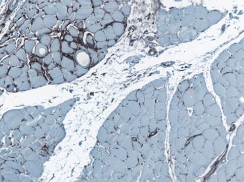
immunohistochemistry (formalin-fixed, paraffin-embedded sections): suitable

isotype

IgG1

shipped in

dry ice

storage temp.

−20°C

target post-translational modification

unmodified

Gene Information

Looking for similar products? Visit Product Comparison Guide

General description

Broadly keratins are grouped into two types on the basis of their biochemical and immunological properties: Type II keratins are a family of basic proteins identified as K1 to K8, whereas type I keratins consists of K9 to K20, belong to the acidic group. It belongs to type I acrylic sub-family and is differentially expressed in various human tissues.
FITC Monoclonal Anti-Cytokeratin Peptide 18 is a purified mouse monoclonal antibody conjugated with fluorescein isothiocyanate (FITC) isomer I. Monoclonal Anti-Cytokeratin Peptide 18 (mouse IgG1 isotype) is derived from the CY-90 hybridoma produced by the fusion of mouse myeloma cells and splenocytes of immunized BALB/c mice. Cytokeratin 18 (CYK18) is a 45 kDa polypeptide, differentially expressed in various human tissues.

Immunogen

human epidermal carcinoma A-431 and MCF-7 human breast cancer cell lines.

Application

Monoclonal Anti-Cytokeratin Peptide 18 FITC antibody produced in mouse has been used in:
  • direct immunofluorescent staining
  • immunohistochemistry
  • double immunofluorescence
  • immunocytochemical and immuno?electron microscopic localization of keratins

Monoclonal Anti-Cytokeratin Peptide 18-FITC antibody:
  • is suitable for immunohistochemistry to identify renal tubular epithelial cells in the extrarenal cells regeneration during acute renal failure
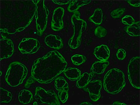
  • is suitable for immunocytochemical and immuno-electron microscopic localisation of keratins in human materno-foetal interaction zone
  • may be used for single or double labeling procedures

Biochem/physiol Actions

Cytokeratins also called keratins (Ks) according to the new nomenclature system.These are known to be the major structural proteins of epithelial cells. It is heteropolymeric in nature i.e. in cells more than one keratin protein is always present. It belongs to type I acrylic sub-family and is differentially expressed in various human tissues. It maintains the structural integrity of extra-embryonic membrane epithelia. Broadly keratins are grouped into two types on the basis of their biochemical and immunological properties: Type II keratins are a family of basic proteins identified as K1 to K8, whereas type I keratins consists of K9 to K20, belong to the acidic group.
Monoclonal anti-cytokeratins are specific markers of epithelial cell differentiation and have been widely used as tools in tumor identification and classification. FITC Monoclonal Anti-Cytokeratin Peptide 18 is a chain specific antibody which can facilitate typing of normal, metaplastic and neoplastic cells. Cytokeratin 18 (CYK18) is a cytoskeletal protein associated with apoptosis. CK-18 is considered as a promising plasma biomarker to identify patients with HELLP (hemolysis, elevated liver enzymes, low platelets) syndrome.

Physical form

Supplied as a solution in 0.01 M phosphate buffered saline, pH 7.4, containing 1% BSA with 15 mM sodium azide as a preservative.

Disclaimer

Unless otherwise stated in our catalog or other company documentation accompanying the product(s), our products are intended for research use only and are not to be used for any other purpose, which includes but is not limited to, unauthorized commercial uses, in vitro diagnostic uses, ex vivo or in vivo therapeutic uses or any type of consumption or application to humans or animals.

Not finding the right product?  

Try our Product Selector Tool.

Storage Class Code

10 - Combustible liquids

Flash Point(F)

Not applicable

Flash Point(C)

Not applicable


Certificates of Analysis (COA)

Search for Certificates of Analysis (COA) by entering the products Lot/Batch Number. Lot and Batch Numbers can be found on a product’s label following the words ‘Lot’ or ‘Batch’.

Already Own This Product?

Find documentation for the products that you have recently purchased in the Document Library.

Visit the Document Library

Cytokeratin 18 is expressed on the hepatocyte plasma membrane surface and interacts with thrombin-antithrombin complexes
Wells MJ, et al.
The Journal of Biological Chemistry, 272(45), 28574-28581 (1997)
Immunofluorescence confocal laser scanning microscopy and immuno-electron microscopic identification of keratins in human materno-foetal interaction zone
Ahenkorah J, et al.
Journal of Cellular and Molecular Medicine, 13(4), 735-748 (2009)
Sofia Batalha et al.
Frontiers in immunology, 14, 1267621-1267621 (2023-11-29)
The clinical prognosis of the HER2-overexpressing (HER2-OE) subtype of breast cancer (BC) is influenced by the immune infiltrate of the tumor. Specifically, monocytic cells, which are promoters of pro-tumoral immunosuppression, and NK cells, whose basal cytotoxic function may be enhanced
Sandeep Gupta et al.
Kidney international, 62(4), 1285-1290 (2002-09-18)
Recovery of renal function following acute tubular necrosis (ATN) is dependent on the replacement of necrotic tubular cells with functional tubular epithelium. The source of these new tubular cells is thought to be resident renal tubular cells. The discovery of
C Lampron et al.
The Journal of biological chemistry, 262(10), 4893-4898 (1987-04-05)
Differentiation of F9 embryonal carcinoma cells by retinoic acid treatment results in extraembryonic endoderm-like cells. The effects of this process on the protein composition of the intermediate filaments were studied by two-dimensional gel electrophoresis and by immunoblotting. By this approach

Our team of scientists has experience in all areas of research including Life Science, Material Science, Chemical Synthesis, Chromatography, Analytical and many others.

Contact Technical Service