Skip to Content
Merck
All Photos(2)

Key Documents

MABF260

Sigma-Aldrich

Anti-NLRC5 Antibody, clone 3H8

clone 3H8, from rat, purified by using protein G

Synonym(s):

Protein NLRC5, Caterpiller protein 16.1, CLR16.1, Nucleotide-binding oligomerization domain protein 27, Nucleotide-binding oligomerization domain protein 4

Sign Into View Organizational & Contract Pricing


About This Item

UNSPSC Code:
12352203
eCl@ss:
32160702
NACRES:
NA.41

biological source

rat

Quality Level

antibody form

purified antibody

antibody product type

primary antibodies

clone

3H8, monoclonal

purified by

using protein G

species reactivity

human

technique(s)

flow cytometry: suitable
immunoprecipitation (IP): suitable
western blot: suitable

isotype

IgG1, kappa

UniProt accession no.

shipped in

wet ice

target post-translational modification

unmodified

Gene Information

human ... NLRC5(84166)

General description

The protein named NLRC5, or also known as Nucleotide-binding oligomerization domain protein 27, Caterpiller protein 16.1 (CLR16.1), or Nucleotide-binding oligomerization domain protein 4 and encoded by the gene NLRC5/NOD27/NOD4, plays a role in the homeostatic control of innate immunity and anti-viral defense mechanisms. NLRC5 also likely regulates the NF kappa B and type 1 interferon signaling response pathways induced upon infection. NLRC5 interacts with CHUK and IKBKB and prevents CHUK and IKBKB phosphorylation thus inhibiting their kinase activity. NLRC5 interacts with DDX58 and IFIH1 proteins and blocks the interaction of MAVS to DDX58 proteins. NLRC5 is localized to the cytoplasm. NLRC5 is expressed in spleen, thymus, lung, brain, tonsil, heart and prostate at moderate levels, but NLRC5 expression is dramatically induced by interferons during pathological infections. Traditional immune responses studies have demonstrated that NLRC5 is a specific and master regulator of major histocompatibility complex (MHC) class I genes as well as related genes involved in MHC class I antigen presentation. The expression of MHC class I genes is regulated by NLRC5 in coordination with the RFX components through an enhanceosome-dependent manner, and the involvement of NLRC5 in MHC class I mediated CD8+ T cell activation, proliferation and cytotoxicity has proved to be critical for host defense against intracellular bacterial infections.

Specificity

Target specificity of clone 3H8 was confirmed by Western blotting using lysates from cells overexpressing exogenously transfected NLRC5 as well as cells knocked down of endogenous NLRC5 by shRNA transfection (Neerincx, A., et al. (2010). J. Biol. Chem. 285(34):26223-26232). Clone 3H8 epitope is present in human NLRC5 spliced isoforms 1 and 4, but absent from isoforms 2, 3, 5, and 6 reported by UniProt (Q86WI3).

Immunogen

BSA-conjugated linear peptide corresponding to the C-terminal sequence of human NLRC5.

Application

Detect NLRC5 using this rat monoclonal antibody, Anti-NLRC5 Antibody, clone 3H8 validated for use in western blotting, IP & Flow Cytometry.
Flow Cytometry Analysis: 0.25 µg from a representative lot detected NLRC5 in Jurkat and Raji cells.

Immunoprecipitation Analysis: A representative lot detected poly(I:C) (Cat. No. 528906) treatment-induced upregulation of endogenous NLRC5 in HeLa cells by IP-Western blotting analysis of both cytosolic and nuclear fractions. A strong NLRC5 nuclear accumulation was seen upon nuclear export inhibition by LepB (Cat. No. 431050) treatment, whereas NLRC5 was mainly cytoplasmic in untreated cells (Neerincx, A., et al. (2012). J. Immunol. 188(10):4940-4950).

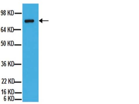
Western Blotting Analysis: A representative lot detected NLRC5 in THP-1 cell lysate as well as exogenously expressed FLAG-tagged NLRC5 in lysate from transfected HEK293T cells. Target band detection was greatly diminished using lysates from NLRC5 siRNA-transfected THP-1 cells. (Neerincx, A., et al. (2010). J. Biol. Chem. 285(34):26223-26232).
Research Category
Inflammation & Immunology
Research Sub Category
Immunoglobulins & Immunology

Quality

Evaluated by Western Blotting in Raji cell lysate.

Western Blotting Analysis: 1 µg/mL of this antibody detected NLRC5 in 10 µg of Raji cell lysate.

Target description

~205 kDa observed. 204.6 kDa (isoform 1) and 201.3 kDa (isoform 4) calculated. Uncharacterized band(s) may appear in some lysates.

Physical form

Protein G Purified
Purified rat monoclonal IgG1κ in buffer containing 0.1 M Tris-Glycine (pH 7.4), 150 mM NaCl with 0.05% sodium azide.

Storage and Stability

Stable for 1 year at 2-8°C from date of receipt.

Other Notes

Concentration: Please refer to the Certificate of Analysis for the lot-specific concentration.

Disclaimer

Unless otherwise stated in our catalog or other company documentation accompanying the product(s), our products are intended for research use only and are not to be used for any other purpose, which includes but is not limited to, unauthorized commercial uses, in vitro diagnostic uses, ex vivo or in vivo therapeutic uses or any type of consumption or application to humans or animals.

Not finding the right product?  

Try our Product Selector Tool.

Storage Class Code

12 - Non Combustible Liquids

WGK

WGK 1

Flash Point(F)

Not applicable

Flash Point(C)

Not applicable


Certificates of Analysis (COA)

Search for Certificates of Analysis (COA) by entering the products Lot/Batch Number. Lot and Batch Numbers can be found on a product’s label following the words ‘Lot’ or ‘Batch’.

Already Own This Product?

Find documentation for the products that you have recently purchased in the Document Library.

Visit the Document Library

Tünde Fekete et al.
Frontiers in immunology, 9, 2314-2314 (2018-10-23)
Unique members of the nucleotide-binding domain leucine-rich repeat (NLR) family have been found to regulate intracellular signaling pathways initiated by other families of pattern recognition receptors (PRR) such as Toll-like receptors (TLRs) and retinoic-acid inducible gene I (RIG-I)-like receptors (RLRs).
Shubhada K Chothe et al.
Frontiers in cellular and infection microbiology, 10, 230-230 (2020-06-09)
Avian influenza viruses (AIVs) cause major economic losses to the global poultry industry. Many host factors have been identified that act as regulators of the inflammatory response and virus replication in influenza A virus (IAV) infected cells including nucleotide-binding oligomerization
Haley E Adcox et al.
mBio, 12(4), e0181621-e0181621 (2021-08-04)
Orientia tsutsugamushi is the etiologic agent of scrub typhus, the deadliest of all diseases caused by obligate intracellular bacteria. Nucleomodulins, bacterial effectors that dysregulate eukaryotic transcription, are being increasingly recognized as key virulence factors. How they translocate into the nucleus
Devin Dersh et al.
Immunity, 54(1), 116-131 (2020-12-04)
Tumors frequently subvert major histocompatibility complex class I (MHC-I) peptide presentation to evade CD8+ T cell immunosurveillance, though how this is accomplished is not always well defined. To identify the global regulatory networks controlling antigen presentation, we employed genome-wide screening in

Our team of scientists has experience in all areas of research including Life Science, Material Science, Chemical Synthesis, Chromatography, Analytical and many others.

Contact Technical Service