Skip to Content
Merck
All Photos(1)

Documents

MAB328

Sigma-Aldrich

Anti-Oligodendrocytes Antibody, clone CE-1

ascites fluid, clone CE-1, Chemicon®

Synonym(s):

MOSP

Sign Into View Organizational & Contract Pricing


About This Item

UNSPSC Code:
12352203
eCl@ss:
32160702
NACRES:
NA.41

biological source

mouse

Quality Level

antibody form

ascites fluid

antibody product type

primary antibodies

clone

CE-1, monoclonal

species reactivity

rat, mouse, chicken, monkey, feline, human

manufacturer/tradename

Chemicon®

technique(s)

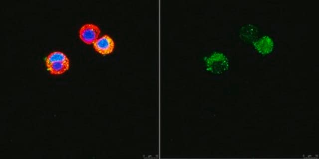
immunocytochemistry: suitable
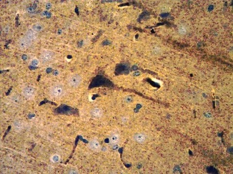
immunohistochemistry (formalin-fixed, paraffin-embedded sections): suitable
immunoprecipitation (IP): suitable

isotype

IgM

suitability

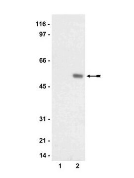
not suitable for Western blot

shipped in

dry ice

target post-translational modification

unmodified

Gene Information

human ... OLIG2(10215)

Specificity

In neonatal rat glia MAB328 stains oligodendrocytes and does not react with type 1 astrocytes, type 2 astrocytes or fibroblasts. Does not stain cultures of rat Schwann cells. In tissue sections MAB328 labels oligodendrocytes and CNS myelin with no labeling of peripheral myelin. MAB328 detects MOSP which is expressed at day 4-5 of neonatal rat, which is about 1-2 after the appearance of GC or sulfatide. The initial expression of MOSP occurs at the stage in development when oligodendrocytes are elaborating processes and just beginning to form membrane sheets (Mu QQ & C Dyer, 1994).

Immunogen

Rat glial membranes and whole brain white matter.

Application

Immunocytochemistry

Immunohistochemistry at 1:1,000-1:5000

Not suggested for use in Western blot.

Optimal working dilutions must be determined by end user.

IMMUNOHISTOCHEMISTRY PROTOCOL FOR MAB328

This antibody has been used successfully on 30 mm, free floating, 4% paraformaldehyde fixed rat brain tissue. All steps are performed under constant agitation. Suggested protocol follows.

1) 3 x 10 minute washes in TBS (with or without 0.25% Triton).

2) Incubate for 30 minutes in TBS with 3% serum (same as host from secondary antibody).

3) Incubate primary antibody diluted appropriately in TBS with 1% serum (same as host from secondary antibody) (with or without 0.25% Triton) for 2 hours at room temperature followed by 16 hours at 4°C.

4) 3 x 10 minute washes in TBS.

5) Incubate with secondary antibody diluted appropriately in TBS with 1% serum (same as host from secondary antibody).

6) 3 x 10 minute washes in TBS.

7) ABC Elite (1:200 Vector Labs) in TBS.

8) 2 x 10 minute washes in TBS.

9) 1 x 10 minute wash in phosphate buffer (no saline).

10) DAB reaction with 0.06% NiCl added for intensification.

11) 2 x 10 minute washes in PBS.

12) 1 x 10 minute wash in phosphate buffer (no saline).
Research Category
Neuroscience
Research Sub Category
Neuronal & Glial Markers
This Anti-Oligodendrocytes Antibody, clone CE-1 is validated for use in IP, IC, IH, IH(P) for the detection of Oligodendrocytes.

Storage and Stability

Maintain at -20°C in undiluted aliquots for up to 12 months. Avoid repeated freeze/thaw cycles.

Legal Information

CHEMICON is a registered trademark of Merck KGaA, Darmstadt, Germany

Disclaimer

Unless otherwise stated in our catalog or other company documentation accompanying the product(s), our products are intended for research use only and are not to be used for any other purpose, which includes but is not limited to, unauthorized commercial uses, in vitro diagnostic uses, ex vivo or in vivo therapeutic uses or any type of consumption or application to humans or animals.

Not finding the right product?  

Try our Product Selector Tool.

Storage Class Code

10 - Combustible liquids

WGK

WGK 1

Flash Point(F)

Not applicable

Flash Point(C)

Not applicable


Certificates of Analysis (COA)

Search for Certificates of Analysis (COA) by entering the products Lot/Batch Number. Lot and Batch Numbers can be found on a product’s label following the words ‘Lot’ or ‘Batch’.

Already Own This Product?

Find documentation for the products that you have recently purchased in the Document Library.

Visit the Document Library

Aleksandra Glavaski-Joksimovic et al.
Cloning and stem cells, 10(1), 75-88 (2008-02-05)
The poor regeneration capability of the mammalian hearing organ has initiated different approaches to enhance its functionality after injury. To evaluate a potential neuronal repair paradigm in the inner ear and cochlear nerve we have previously used embryonic neuronal tissue
Nicole M Jones et al.
Journal of neurochemistry, 89(1), 157-167 (2004-03-20)
Hypoxic preconditioning (HP) 24 h before hypoxic-ischemic (HI) injury confers significant neuroprotection in neonatal rat brain. Recent studies have shown that the mitogen-activated protein kinase (MAPK) and phosphatidylinositol-3-kinase (PI3K) intracellular signaling pathways play a role in the induction of tolerance
Adela Quesada et al.
Journal of neuroscience research, 89(5), 674-688 (2011-02-22)
The retina of nonmammalian vertebrates has a loose myelin that enwraps the large axons of the ganglion cells in all areas, whereas that of mammals lacks myelin, with some exceptions, such as the rabbit retina, which shows compact myelin restricted
Ying Ding et al.
BMC neuroscience, 10, 35-35 (2009-04-21)
Bone marrow mesenchymal stem cells (MSCs) are one of the potential tools for treatment of the spinal cord injury; however, the survival and differentiation of MSCs in an injured spinal cord still need to be improved. In the present study
Dheeraj Pelluru et al.
The European journal of neuroscience, 43(10), 1298-1306 (2015-09-16)
A distributed network of neurons regulates wake, non-rapid eye movement (NREM) sleep, and REM sleep. However, there are also glia in the brain, and there is growing evidence that neurons and astroglia communicate intimately to regulate behaviour. To identify the

Our team of scientists has experience in all areas of research including Life Science, Material Science, Chemical Synthesis, Chromatography, Analytical and many others.

Contact Technical Service