17-10394
Rac1/cdc42 Activation Magnetic Beads Pulldown Assay
The Rac1/cdc42 Activation Magnetic Beads Pulldown Assay provides an effective method for detecting Rac & Cdc42 activity in cell lysates with higher yield & easier process utilizing magenetic bead properties.
Sign Into View Organizational & Contract Pricing
All Photos(2)
About This Item
UNSPSC Code:
12161503
eCl@ss:
32161000
NACRES:
NA.32
Recommended Products
Quality Level
species reactivity
human, rat, mouse
manufacturer/tradename
Upstate®
technique(s)
activity assay: suitable
affinity binding assay: suitable (G-protein)
NCBI accession no.
UniProt accession no.
shipped in
dry ice
Gene Information
human ... RAC1(5879)
General description
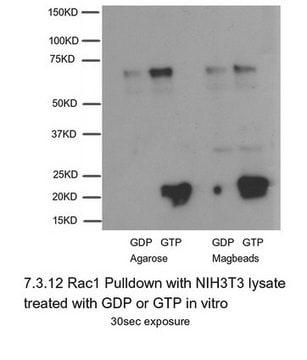
Rac1/cdc42 Activation Magnetic Beads Pulldown Assay (Cat. No. 17-10394) provides an effective method for detecting Rac and Cdc42 activity in cell lysates. This assay uses the downstream effector of Rac/Cdc42, p21-activated protein kinase (PAK1), to isolated the active GTP-bound form of Rac/Cdc42 from the sample. The p21 binding domain (PBD) of PAK1 is expressed as a GST-fusion protein and coupled to magnetic beads. After magnetic rack separation, an immunoblot is performed and the activated Rac/Cdc42 is detected with specific monoclonal antibodies, followed by HRP-conjugated secondary antibody and ECL reagent.
Specificity
Species Cross-reactivity: Human and mouse. Predicted to cross-react with all mammalian species.
Anti-Rac1, clone 23A8: Species Cross-reactivity: Human, mouse and rat. Other species cross-reactivity unknown.
Anti-cdc42 Species Cross-reactivity: Human, mouse, rat and dog. Other species cross-reactivity unknown.
Anti-Rac1, clone 23A8: Species Cross-reactivity: Human, mouse and rat. Other species cross-reactivity unknown.
Anti-cdc42 Species Cross-reactivity: Human, mouse, rat and dog. Other species cross-reactivity unknown.
Application
Research Category
Cell Signaling
Signaling
Cell Signaling
Signaling
Components
Rac1/cdc42 Assay Reagent (Pak-1PBD, Magnetic beads): 300 ug of Pak-1 PBD in 150 uL of glutathione magnetic beads, provided as a 50% beads slurry in PBS containing 50% glycerol for a final volume of 300 uL.
Anti-Rac1, clone 23A8: One vial containing 250 ug of purified IgG2akappa in 250 uL of storage buffer (0.1 M Tris-glycine, pH 7.4, 0.15 M NaCl, containing 0.05% sodium azide).
Anti-cdc42, mouse monoclonal IgG1: One vial containing 50 ug of purified mouse IgG1 in 200 uL of 10 mM sodium phosphate, pH 7.5, 75 mM NaCl, 0.75 mM sodium azide, 0.5 mg/mL BSA and 50% glycerol.
Mg2+ Lysis/Wash Buffer, 5X, Catalog # 20-168 : Two vials, each vial containing 18 mL of 5X MLB: 125 mM HEPES, pH 7.5, 750 mM NaCl, 5% Igepal CA-630, 50 mM MgCl2, 5 mM EDTA and 10% glycerol.
100X GTPγS, 10mM, Catalog # 20-176: One vial containing 50 μL of 10 mM GTPγS, 100X stock, in 50 mM Tris-HCl, pH 7.8, non-hydrolyzable analog of GTP. Sufficient to label 5 mL of cell lysates.
100X GDP, 100mM, Catalog # 20-177: One vial containing 50 μL of 100 mM GDP, 100X stock, in 50 mM Tris-HCl, pH 7.8. GDP (Guanosine 5′-Diphosphate) for in vitro labeling of G-proteins in the inactive form. Sufficient to label 5ml of cell lysates.
Quality
Routinely evaluated by precipitating Rac1-GTP and cdc42-GTP from 3T3/A31 and HeLa cell lysates that had been loaded with GTPγS(lysates were loaded in vitro with GTPγS for cdc42-GTP). The precipitated Rac1-GTP and cdc42-GTP was detected by immunoblot analysis using anti-Rac1 (1 μg/ml).
Storage and Stability
1 year at -20°C from date of shipment
Legal Information
UPSTATE is a registered trademark of Merck KGaA, Darmstadt, Germany
Disclaimer
Unless otherwise stated in our catalog or other company documentation accompanying the product(s), our products are intended for research use only and are not to be used for any other purpose, which includes but is not limited to, unauthorized commercial uses, in vitro diagnostic uses, ex vivo or in vivo therapeutic uses or any type of consumption or application to humans or animals.
Signal Word
Danger
Hazard Statements
Precautionary Statements
Hazard Classifications
Aquatic Acute 1 - Aquatic Chronic 2 - Eye Dam. 1
Storage Class Code
10 - Combustible liquids
Certificates of Analysis (COA)
Search for Certificates of Analysis (COA) by entering the products Lot/Batch Number. Lot and Batch Numbers can be found on a product’s label following the words ‘Lot’ or ‘Batch’.
Already Own This Product?
Find documentation for the products that you have recently purchased in the Document Library.
Qiong Xu et al.
Experimental cell research, 390(1), 111933-111933 (2020-03-07)
Current studies have shown that POTE ankyrin domain family members have high expressions as tumor antigens in malignant tumors, such as prostate cancer, ovarian cancer, breast cancer and the like. POTEE is a member of the POTE anchor protein family
Hong Li et al.
The Journal of clinical investigation, 131(22) (2021-10-01)
Epoxyeicosatrienoic acids (EETs) have potent antiinflammatory properties. Hydrolysis of EETs by soluble epoxide hydrolase/ epoxide hydrolase 2 (sEH/EPHX2) to less active diols attenuates their antiinflammatory effects. Macrophage activation is critical to many inflammatory responses; however, the role of EETs and
Hooi Ting Hu et al.
STAR protocols, 2(3), 100625-100625 (2021-07-06)
Extracellular vesicles (EVs) play important roles in extracellular trafficking and signaling. Here, we separate EVs by differential centrifugation. EVs separated by this approach are called large EVs (l-EVs) and small EVs (s-EVs), reflecting particle size, which sediment based on different
Jie Li et al.
Scientific reports, 6, 26971-26971 (2016-05-28)
Hepatocellular carcinoma (HCC) is complicated by aggressive migration and invasion, which contribute to the increased mortality of HCC patients. The NKD1 protein is abnormally expressed in many neoplasms and plays an important role in tumor progression. However, the regulation and
Jessica Diring et al.
Nature cell biology, 21(7), 845-855 (2019-06-19)
RPEL proteins, which contain the G-actin-binding RPEL motif, coordinate cytoskeletal processes with actin dynamics. We show that the ArhGAP12- and ArhGAP32-family GTPase-activating proteins (GAPs) are RPEL proteins. We determine the structure of the ArhGAP12/G-actin complex, and show that G-actin contacts
Our team of scientists has experience in all areas of research including Life Science, Material Science, Chemical Synthesis, Chromatography, Analytical and many others.
Contact Technical Service
