Skip to Content
Merck
All Photos(1)

Key Documents

R5403

Sigma-Aldrich

Monoclonal Anti-Rhodopsin antibody produced in mouse

clone 1D4, purified immunoglobulin, buffered aqueous solution

Synonym(s):

Anti-CSNBAD1, Anti-OPN2, Anti-RP4

Sign Into View Organizational & Contract Pricing


About This Item

MDL number:
UNSPSC Code:
12352203
NACRES:
NA.41

biological source

mouse

Quality Level

conjugate

unconjugated

antibody form

purified immunoglobulin

antibody product type

primary antibodies

clone

1D4, monoclonal

form

buffered aqueous solution

species reactivity

human, rat, bovine

technique(s)

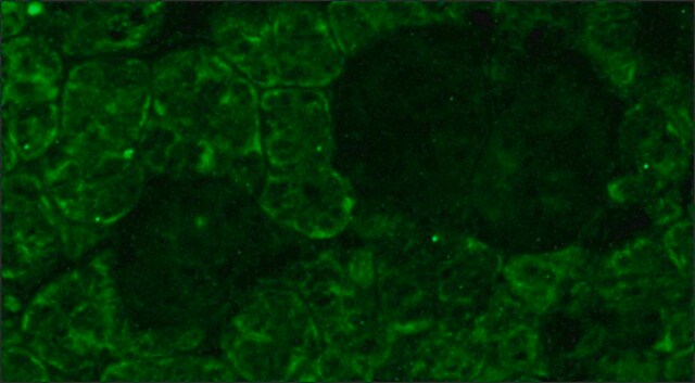
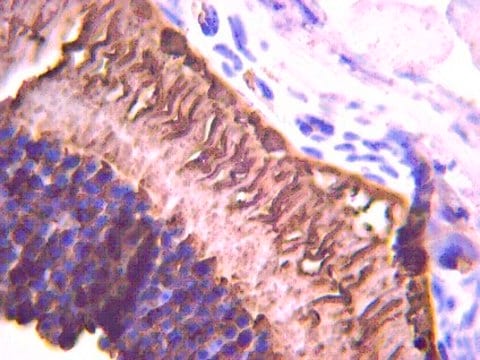
immunocytochemistry: 1:1,000 using human retinal samples
western blot: 1:1,000 using Sf9 cells expressing the bovine gene

isotype

IgG1

UniProt accession no.

shipped in

wet ice

storage temp.

−20°C

target post-translational modification

unmodified

Gene Information

human ... RHO(6010)
rat ... Rho(24717)

General description

Vision involves the conversion of light into electrochemical signals that are processed by the retina and subsequently sent to and interpreted by the brain. The process of converting light to an electrochemical signal begins when the membrane-bound protein, rhodopsin, absorbs light within the retina. Photoexcitation of rhodopsin causes the cytoplasmic surface of the protein to become catalytically active. In the active state, rhodopsin activates transducin, a GTP binding protein. Once activated, transducin promotes the hydrolysis of cGMP by phosphodiesterase (PDE). The decrease of intracellular cGMP concentrations causes the ion channels within the outer segment of the rod or cone to close, thus causing membrane hyperpolarization and, eventually, signal transmission. Rhodopsin′s activity is believed to be shut off by its phosphorylation followed by binding of the soluble protein arrestin.

Specificity

By Western blot, mouse monoclonal clone 1D4 anti-Rhodopsin antibody detects an ~40 kDa protein representing rhodopsin from Sf 9 cells expressing the bovine gene. Immunocytochemical staining of rhodopsin in human retinal samples results in staining of both rod and cone outer segments.
The epitope for this antibody has been localized to the C-terminal nine amino acids of bovine rhodopsin known as the 1D4 epitope, and detects rhodopsin from human and bovine retinal samples.

Immunogen

bovine rhodopsin.

Application

Applications in which this antibody has been used successfully, and the associated peer-reviewed papers, are given below.
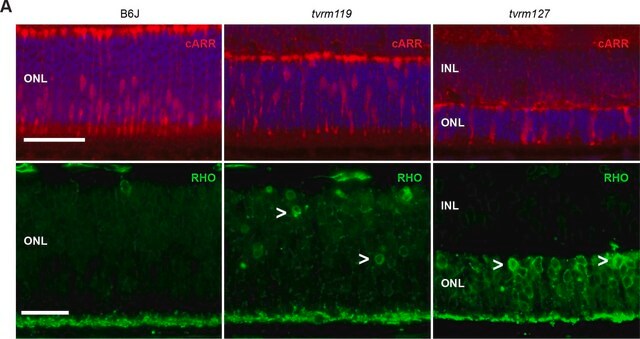
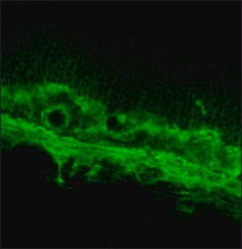
Immunohistochemistry (1 paper)
Mouse monoclonal clone 1D4 anti-Rhodopsin antibody has been successfully used in Western blot, immunocytochemistry, and immunoprecipitation. It is used as a probe to determine the presence and roles of rhodopsin in the conversion of light into electrochemical signals.

Physical form

Solution in phosphate buffered saline containing 1 mg/mL BSA and 0.05% sodium azide.

Disclaimer

Unless otherwise stated in our catalog or other company documentation accompanying the product(s), our products are intended for research use only and are not to be used for any other purpose, which includes but is not limited to, unauthorized commercial uses, in vitro diagnostic uses, ex vivo or in vivo therapeutic uses or any type of consumption or application to humans or animals.

Not finding the right product?  

Try our Product Selector Tool.

Storage Class Code

10 - Combustible liquids

WGK

nwg

Flash Point(F)

Not applicable

Flash Point(C)

Not applicable


Certificates of Analysis (COA)

Search for Certificates of Analysis (COA) by entering the products Lot/Batch Number. Lot and Batch Numbers can be found on a product’s label following the words ‘Lot’ or ‘Batch’.

Already Own This Product?

Find documentation for the products that you have recently purchased in the Document Library.

Visit the Document Library

Angelo Felline et al.
Computational and structural biotechnology journal, 19, 6020-6038 (2021-12-02)
Failure of a protein to achieve its functional structural state and normal cellular location contributes to the etiology and pathology of heritable human conformational diseases. The autosomal dominant form of retinitis pigmentosa (adRP) is an incurable blindness largely linked to
Maria Carmela Latella et al.
Molecular therapy. Nucleic acids, 5(11), e389-e389 (2016-11-23)
The bacterial CRISPR/Cas system has proven to be an efficient tool for genetic manipulation in various organisms. Here we show the application of CRISPR-Cas9 technology to edit the human Rhodopsin (RHO) gene in a mouse model for autosomal dominant Retinitis
Jun Yin et al.
Investigative ophthalmology & visual science, 53(8), 4943-4951 (2012-06-30)
In experimental eye research, zebrafish has become a powerful model for human retina disorders. The purpose of the present study is the characterization of antibodies commonly employed in zebrafish models for rod photoreceptor degeneration. The 1D4 monoclonal antibody, developed against
Silvia Ravera et al.
Cells, 12(17) (2023-09-08)
Dysfunction of the retinal pigment epithelium (RPE) is associated with several diseases characterized by retinal degeneration, such as diabetic retinopathy (DR). However, it has recently been proposed that outer retinal neurons also participate in the damage triggering. Therefore, we have
Kirk Haltaufderhyde et al.
Photochemistry and photobiology, 91(1), 117-123 (2014-10-01)
Human skin is constantly exposed to solar light containing visible and ultraviolet radiation (UVR), a powerful skin carcinogen. UVR elicits cellular responses in epidermal cells via several mechanisms: direct absorption of short-wavelength UVR photons by DNA, oxidative damage caused by

Our team of scientists has experience in all areas of research including Life Science, Material Science, Chemical Synthesis, Chromatography, Analytical and many others.

Contact Technical Service