Skip to Content
Merck

HPA001562

Sigma-Aldrich

Anti-ATF3 antibody produced in rabbit

enhanced validation

Prestige Antibodies® Powered by Atlas Antibodies, affinity isolated antibody, buffered aqueous glycerol solution

Synonym(s):

ATF3 Antibody - Anti-ATF3 antibody produced in rabbit, Atf3 Antibody, Anti-Activating transcription factor 3 antibody produced in rabbit, Anti-Cyclic AMP-dependent transcription factor ATF-3 antibody produced in rabbit

Sign Into View Organizational & Contract Pricing


About This Item

MDL number:
UNSPSC Code:
12352203
Human Protein Atlas Number:
NACRES:
NA.43

Pricing and availability is not currently available.

biological source

rabbit

Quality Level

conjugate

unconjugated

antibody form

affinity isolated antibody

antibody product type

primary antibodies

clone

polyclonal

product line

Prestige Antibodies® Powered by Atlas Antibodies

form

buffered aqueous glycerol solution

species reactivity

human

enhanced validation

orthogonal RNAseq
Learn more about Antibody Enhanced Validation

General description

Activating transcription factor 3 (ATF3) gene is mapped to human chromosome 1q32.3. The encoded protein belongs to the cAMP-response element binding (CREB) protein family.
Rabbit polyclonal anti-ATF3 antibody reacts with human activating transcription factor 3.

Immunogen

Cyclic AMP-dependent transcription factor ATF-3 recombinant protein epitope signature tag (PrEST)

Application

Anti-ATF3 antibody produced in rabbit has been used in:
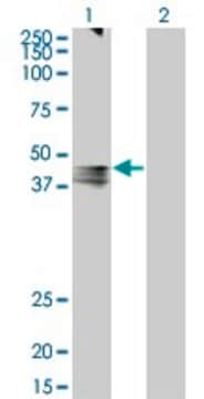
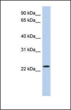
  • western blotting
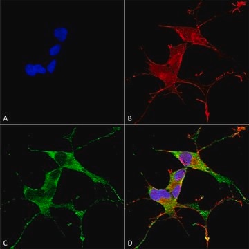
  • immunofluorescence
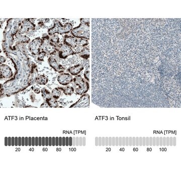
  • immunohistochemical staining
  • chromatin immunoprecipitation (ChIP)

Biochem/physiol Actions

Activating transcription factor 3 (ATF3), a cyclic AMP-dependent transcription factor stimulates transcription by sequestering inhibitory cofactors. ATF3 responds to stress signals and is involved in the regulation of immune and metabolic homeostasis. It helps to prevent chronic inflammation and starvation responses. ATF3 expression is induced in response to eye injury. It serves as a marker for neuronal injury.

Features and Benefits

Prestige Antibodies® are highly characterized and extensively validated antibodies with the added benefit of all available characterization data for each target being accessible via the Human Protein Atlas portal linked just below the product name at the top of this page. The uniqueness and low cross-reactivity of the Prestige Antibodies® to other proteins are due to a thorough selection of antigen regions, affinity purification, and stringent selection. Prestige antigen controls are available for every corresponding Prestige Antibody and can be found in the linkage section.

Every Prestige Antibody is tested in the following ways:
  • IHC tissue array of 44 normal human tissues and 20 of the most common cancer type tissues.
  • Protein array of 364 human recombinant protein fragments.

Linkage

Corresponding Antigen APREST77475

Physical form

Solution in phosphate-buffered saline, pH 7.2, containing 40% glycerol and 0.02% sodium azide

Legal Information

Prestige Antibodies is a registered trademark of Merck KGaA, Darmstadt, Germany

Disclaimer

Unless otherwise stated in our catalog or other company documentation accompanying the product(s), our products are intended for research use only and are not to be used for any other purpose, which includes but is not limited to, unauthorized commercial uses, in vitro diagnostic uses, ex vivo or in vivo therapeutic uses or any type of consumption or application to humans or animals.

Not finding the right product?  

Try our Product Selector Tool.

Storage Class Code

10 - Combustible liquids

WGK

WGK 1

Personal Protective Equipment

dust mask type N95 (US), Eyeshields, Gloves

Certificates of Analysis (COA)

Search for Certificates of Analysis (COA) by entering the products Lot/Batch Number. Lot and Batch Numbers can be found on a product’s label following the words ‘Lot’ or ‘Batch’.

Need A Sample COA?

This is a sample Certificate of Analysis (COA) and may not represent a recently manufactured lot of this specific product.

Already Own This Product?

Find documentation for the products that you have recently purchased in the Document Library.

Visit the Document Library

Saisai Wei et al.
The Journal of biological chemistry, 289(13), 8947-8959 (2014-02-21)
Mutant p53 proteins (mutp53) often acquire oncogenic activities, conferring drug resistance and/or promoting cancer cell migration and invasion. Although it has been well established that such a gain of function is mainly achieved through interaction with transcriptional regulators, thereby modulating
Inducible ATF3-NFAT axis aggravates podocyte injury
Zhang H, et al.
Journal of Molecular Medicine, 96(1), 53-64 (2018)
Activating transcription factor 3 (ATF3) expression in the neural retina and optic nerve of zebrafish during optic nerve regeneration
Saul KE, et al.
Comparative Biochemistry and Physiology. Part A, Molecular & Integrative Physiology, 155(2), 172-182 (2010)
Makoto Edagawa et al.
The Journal of biological chemistry, 289(31), 21544-21561 (2014-06-19)
Death receptor 5 (DR5) is a death domain-containing transmembrane receptor that triggers cell death upon binding to its ligand, TNF-related apoptosis-inducing ligand (TRAIL), and a combination of TRAIL and agents that increase the expression of DR5 is expected to be
Activating transcription factor 3 attenuates chemokine and cytokine expression in mouse skeletal muscle after exercise and facilitates molecular adaptation to endurance training
Fernandez VR, et al.
Faseb Journal, 31(2), 840-851 (2016)

Our team of scientists has experience in all areas of research including Life Science, Material Science, Chemical Synthesis, Chromatography, Analytical and many others.

Contact Technical Service