A3429
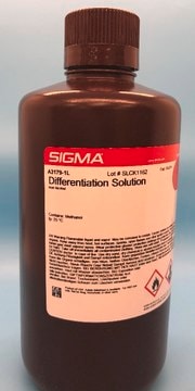
Differentiation Solution
for the differentiation of regressive hematoxylin stains
Synonym(s):
Acid Alcohol
Sign Into View Organizational & Contract Pricing
All Photos(2)
About This Item
Recommended Products
Quality Level
form
solution
shelf life
40 mo.
color
colorless
application(s)
hematology
histology
storage temp.
room temp
Looking for similar products? Visit Product Comparison Guide
General description
Differentiation solution comprises acidified ethanol or isopropanol that is used to decolorize hematoxylin stain. The duration of exposure to the solution determines the degree of decolorization. Differentiation is a crucial step in hematoxylin-eosin staining to achieve the proper color contrast necessary for visualization, by selectively removing excess hematoxylin from the tissue section and leaving behind the desired staining intensity.
Application
Differentiation solution is employed mainly for the differentiation of regressive hematoxylin stains. It has been used in the following studies:
- the prediction of pulmonary metastasis progression in osteosarcoma patients
- the development of multispectral imaging to detect melanin
Acidified alcohol solution for the differentiation of regressive hematoxylin stains.
Features and Benefits
- Desired staining patterns can be obtained by varying the degree of exposure to the differentiation solution.
- Results are sharper and more rapid compared to other differentiation methods.
- Crisp and clear differentiation between cellular structures is obtainable when performed according to Sigma-Aldrich Procedure No. GHS.
Principle
In regressive hematoxylin staining, a highly concentrated hematoxylin solution is used to rapidly overstain both nuclei and cytoplasm, and the cytoplasm is subsequently decolorized and excess hematoxylin is removed from the nucleus with dilute acid. Improper differentiation leaves excess residual hematoxylin that obscures the fine details of nuclear structures and chromatin and prevents eosin uptake.
Signal Word
Danger
Hazard Statements
Precautionary Statements
Hazard Classifications
Eye Irrit. 2 - Flam. Liq. 2 - Met. Corr. 1 - STOT SE 2
Target Organs
Eyes,Central nervous system
Storage Class Code
3 - Flammable liquids
WGK
WGK 1
Flash Point(F)
71.6 °F
Flash Point(C)
22 °C
Certificates of Analysis (COA)
Search for Certificates of Analysis (COA) by entering the products Lot/Batch Number. Lot and Batch Numbers can be found on a product’s label following the words ‘Lot’ or ‘Batch’.
Already Own This Product?
Find documentation for the products that you have recently purchased in the Document Library.
M Jesús Andrés-Manzano et al.
Methods in molecular biology (Clifton, N.J.), 1339, 85-99 (2015-10-09)
Methods for staining tissues with Oil Red O and hematoxylin-eosin are classical histological techniques that are widely used to quantify atherosclerotic burden in mouse tissues because of their ease of use, reliability, and the large amount of information they provide.
Nuclear staining with alum hematoxylin
B D Llewellyn
Biotechnic & Histochemistry, 84(9), 159-177 (2009)
Oil Red O and Hematoxylin and Eosin Staining for Quantification of Atherosclerosis Burden in Mouse Aorta and Aortic Root
Andres-Manzano, et al.
Methods in Molecular Biology, 1339 (2015)
Pia M Teleman et al.
Acta obstetricia et gynecologica Scandinavica, 88(8), 927-932 (2009-07-07)
To outline possible associations between urinary incontinence (UI) and serum levels of steroid hormones in middle-aged women. Community-based observational study. All women aged 50-59 living in the Lund area by December 1995 were invited to a screening procedure. Sixty-four percent
Multispectral imaging as a tool for melanin detection
Kalleberg, et al.
Journal of Histotechnology, 38(1), 14-21 (2015)
Our team of scientists has experience in all areas of research including Life Science, Material Science, Chemical Synthesis, Chromatography, Analytical and many others.
Contact Technical Service


