Skip to Content
Merck
All Photos(2)

Key Documents

ABS535

Sigma-Aldrich

Anti-Arginase-1 Antibody

from chicken

Synonym(s):

Type 1 Arginase

ARG1

Sign Into View Organizational & Contract Pricing


About This Item

UNSPSC Code:
12352203
eCl@ss:
32160702
NACRES:
NA.41

biological source

chicken

Quality Level

antibody form

purified immunoglobulin

antibody product type

primary antibodies

clone

polyclonal

species reactivity

guinea pig, human, mouse, rat

technique(s)

immunohistochemistry: suitable
western blot: suitable

isotype

IgY

NCBI accession no.

UniProt accession no.

shipped in

wet ice

target post-translational modification

unmodified

Gene Information

human ... ARG1(383)
mouse ... Arg1(11846)
rat ... Arg1(29221)

General description

Arginase-1, the protein encoded by the ARG1 gene, is the enzyme that catalyzes the breakdown of L-arginine into ornithine and urea. ARG1 is a key player in the Urea cycle. There are two distinct isoforms of Arginase, Arginase I and Arginase II. Arginase I (also called the liver type arginase or Type 1 arginase) is a highly expressed cytosolic enzyme in the liver and other tissues, including the brain. Arginase II or kidney-type arginase is also widely expressed at low levels in many tissues but localized to the mitochondria primarily. Interestingly, Arginase 1 has neuroprotective properties. Arginase I is essential for protecting motor neurons from nitric oxide dependent apoptosis that can be induced upon cellular stress or trophic factor removal. By suppressing nitric oxide production, Arginase prevents the formation of peroxynitrite and its subsequent cellular toxicity. Furthermore, Arginase 1 appears to play critical roles throughout the CNS for survival and regeneration of neurons and axons. In cancer, Arginase 1, already a useful marker for hepatocytes is also an excellent marker for hepatocellular cancers.

Immunogen

Full-length recombinant protein corresponding to Rat Arginase-1.

Application

Immunohistochemistry Analysis: A representative lot detected Arginase-1 in bronchoalveolar lavage cells (Zimmermann, N., et al. (2003). J. Clin. Invest. 111:1863–1874 (2003)).
Western Blotting Analysis: A representative lot detected Arginase-1 in RAW cell lysate (Morris, S., et al. (1998). Am J Physiol Endocrinol Metab 275:E740-E747, 1998).
Research Category
Signaling
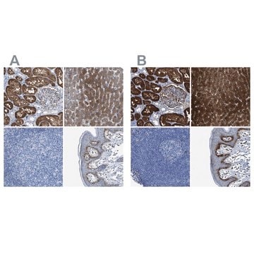
This Anti-Arginase-1 Antibody is validated for use in Western Blotting and Immunohistochemistry for the detection of Arginase-1.

Quality

Evaluated by Western Blotting in mouse LN tissue lysate.

Western Blotting Analysis: 2.0 µg/mL of this antibody detected Arginase-1 in 10 µg of mouse LN tissue lysate.

Target description

~ 38 kDa observed

Physical form

Format: Purified
IgY purified from Yolk
Purified chicken Polyclonal other in buffer containing PBS with 0.05% sodium azide.

Storage and Stability

Stable for 1 year at 2-8°C from date of receipt.

Other Notes

Concentration: Please refer to lot specific datasheet.

Disclaimer

Unless otherwise stated in our catalog or other company documentation accompanying the product(s), our products are intended for research use only and are not to be used for any other purpose, which includes but is not limited to, unauthorized commercial uses, in vitro diagnostic uses, ex vivo or in vivo therapeutic uses or any type of consumption or application to humans or animals.

Not finding the right product?  

Try our Product Selector Tool.

Storage Class Code

12 - Non Combustible Liquids

WGK

WGK 2

Flash Point(F)

Not applicable

Flash Point(C)

Not applicable


Certificates of Analysis (COA)

Search for Certificates of Analysis (COA) by entering the products Lot/Batch Number. Lot and Batch Numbers can be found on a product’s label following the words ‘Lot’ or ‘Batch’.

Already Own This Product?

Find documentation for the products that you have recently purchased in the Document Library.

Visit the Document Library

Tim Tian Y Han et al.
Frontiers in bioengineering and biotechnology, 9, 642465-642465 (2021-04-06)
Cell-based therapies involving the delivery of adipose-derived stromal cells (ASCs) on decellularized adipose tissue (DAT) scaffolds are a promising approach for soft tissue augmentation and reconstruction. Our lab has recently shown that culturing human ASCs on DAT scaffolds within a
Qianru Yang et al.
Biomaterials, 276, 121060-121060 (2021-08-23)
Our understanding of biomaterials in the brain have been greatly enhanced by advancements in in vivo imaging technologies such as two-photon microscopy. However, when applied to chronic studies, two-photon microscopy enables high-resolution imaging only in superficial regions due to inflammatory
Daniella Bianchi-Frias et al.
Molecular cancer research : MCR, 17(1), 321-331 (2018-09-19)
The incidence of prostate cancer is directly linked to age, but age-associated changes that facilitate prostate cancer development and progression are poorly understood. This study investigated age-related changes in the prostate microenvironment for their influence on prostate cancer behavior. Prostate
X Sally Zheng et al.
Acta biomaterialia, 103, 81-91 (2019-12-22)
Electrical stimulation of the muscle has been proven efficacious in preventing atrophy and/or reanimating paralyzed muscles. Intramuscular electrodes made from metals have significantly higher Young's Moduli than the muscle tissues, which has the potential to cause chronic inflammation and decrease
Asiyeh Golabchi et al.
Biomaterials, 180, 225-239 (2018-07-28)
The chronic performance of implantable neural electrodes is hindered by inflammatory brain tissue responses, including microglia activation, glial scarring, and neuronal loss. Melatonin (MT) has shown remarkable neuroprotective and neurorestorative effects in treating central nervous system (CNS) injuries and degeneration

Our team of scientists has experience in all areas of research including Life Science, Material Science, Chemical Synthesis, Chromatography, Analytical and many others.

Contact Technical Service