Skip to Content
Merck
All Photos(3)

Documents

C4731

Sigma-Aldrich

Anti-Calnexin antibody produced in rabbit

enhanced validation

IgG fraction of antiserum, buffered aqueous solution

Synonym(s):

Anti-CNX, Anti-IP90, Anti-P90

Sign Into View Organizational & Contract Pricing


About This Item

MDL number:
UNSPSC Code:
12352203
NACRES:
NA.44

biological source

rabbit

Quality Level

conjugate

unconjugated

antibody form

IgG fraction of antiserum

antibody product type

primary antibodies

clone

polyclonal

form

buffered aqueous solution

mol wt

antigen 90 kDa

species reactivity

canine, rat, mouse, human

packaging

antibody small pack of 25 μL

enhanced validation

independent
Learn more about Antibody Enhanced Validation

technique(s)

immunoprecipitation (IP): 1:2,000 using whole cell RIPA lysate of the human epitheloid carcinoma HeLa cell line
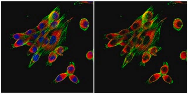
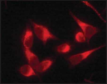
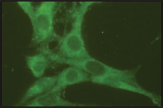
indirect immunofluorescence: 1:200 using 3% paraformaldehyde-fixed, 0.5% Triton X-100 treated, Madin Darby canine kidney MDCK cell line
microarray: suitable
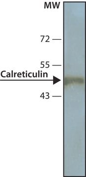
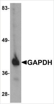
western blot: 1:2,000 using whole cell RIPA lysate of the human hepatocytoma HepG2 cell line

UniProt accession no.

shipped in

dry ice

storage temp.

−20°C

target post-translational modification

unmodified

Gene Information

human ... CANX(821)
mouse ... Canx(12330)
rat ... Canx(29144)

General description

Calnexin contains a long (461 amino acids) N-terminal Ca2+ -binding domain extending into the lumen of the endoplasmic reticulum (ER), a short (22 amino acids) transmembrane segment and an acidic cytosolic domain (96 amino acids).
Calnexin, is a calcium binding type I integral membrane protein present mainly in the endoplasmic reticulum. It plays a pivotal role in quality control processes during protein synthesis and folding. Anti-calnexin antibody (diluted 1: 300) can be used in dual immunofluorescence staining to visualize TG2 transport.

Specificity

Rabbit anti-calnexin antibody reacts specifically with calnexin (90 kDa) of dog, mouse, rat and human.

Immunogen

Synthetic peptide corresponding to the c-terminus of human calnexin (amino acids 573-592) conjugated to KLH.

Application

Anti-Calnexin antibody produced in rabbit has been used in:
  • western blot analysis
  • immunofluorescence
  • dual immunofluorescence staining

Biochem/physiol Actions

Calnexin binds newly synthesized glycoproteins and misfolded proteins and is believed to play a critical role in quality control processes during protein synthesis and folding. Calnexin acts as a lectin-like chaperone that binds oligosaccharide residues of newly synthesized N-linked glycoproteins. Calnexin has been shown to be associated with several cell surface proteins, including major histocompatibility complex (MHC) class I heavy chain, T-cell receptor (TCR), and B cell membrane immunoglobulin during translocation through the endoplasmic reticulum (ER). It also forms complexes with other resident ER proteins involved in Ca2+ -dependent retention of proteins.

Physical form

Solution in 0.01 M phosphate buffered saline, pH 7.4, containing 15 mM sodium azide.

Disclaimer

Unless otherwise stated in our catalog or other company documentation accompanying the product(s), our products are intended for research use only and are not to be used for any other purpose, which includes but is not limited to, unauthorized commercial uses, in vitro diagnostic uses, ex vivo or in vivo therapeutic uses or any type of consumption or application to humans or animals.

Not finding the right product?  

Try our Product Selector Tool.

Storage Class Code

10 - Combustible liquids

WGK

WGK 2


Certificates of Analysis (COA)

Search for Certificates of Analysis (COA) by entering the products Lot/Batch Number. Lot and Batch Numbers can be found on a product’s label following the words ‘Lot’ or ‘Batch’.

Already Own This Product?

Find documentation for the products that you have recently purchased in the Document Library.

Visit the Document Library

Calnexin and calreticulin, molecular chaperones of the endoplasmic reticulum
Calreticulin, 49-62 (2003)
Quality control system of the endoplasmic reticulum and related diseases
Wu JC, et al.
Acta biochimica et biophysica Sinica, 38(4), 219-226 (2006)
Markus Düchler et al.
Cells, 8(7) (2019-07-05)
Cancer-induced immunosuppression is antigen-specific rather than systemic and the mechanisms for the antigen specificity are incompletely understood. Here we explore the option that tumor-associated antigens (TAAs) may be transferred to antigen-presenting cells (APCs), together with immunosuppressive molecules, through cancer-derived small
Valeria Uribe et al.
Human molecular genetics, 21(9), 1954-1967 (2012-01-21)
Apoptosis, or programmed cell death, is a cellular pathway involved in normal cell turnover, developmental tissue remodeling, embryonic development, cellular homeostasis maintenance and chemical-induced cell death. Caspases are a family of intracellular proteases that play a key role in apoptosis.
Gokhan Yildiz et al.
PloS one, 8(5), e64016-e64016 (2013-05-22)
Senescence is a permanent proliferation arrest in response to cell stress such as DNA damage. It contributes strongly to tissue aging and serves as a major barrier against tumor development. Most tumor cells are believed to bypass the senescence barrier

Our team of scientists has experience in all areas of research including Life Science, Material Science, Chemical Synthesis, Chromatography, Analytical and many others.

Contact Technical Service