MABC969
Anti-PD-L2 Antibody, clone 24F.10C12
clone 24F.10C12, from mouse
Synonym(s):
Programmed cell death 1 ligand 2, B7-DC, B7 dendritic cell molecule, B7-H2, Butyrophilin B7-DC, CD273, PD-1 ligand 2, PD-L2, PDCD1 ligand 2, Programmed death ligand 2
About This Item
Recommended Products
biological source
mouse
Quality Level
antibody form
purified immunoglobulin
antibody product type
primary antibodies
clone
24F.10C12, monoclonal
species reactivity
human
technique(s)
flow cytometry: suitable
immunohistochemistry: suitable
neutralization: suitable
western blot: suitable
isotype
IgG2aκ
NCBI accession no.
UniProt accession no.
shipped in
dry ice
target post-translational modification
unmodified
Gene Information
human ... PDCD1LG2(80380)
Related Categories
General description
Specificity
Immunogen
Application
Flow Cytometry Analysis: A representative lot immunostained the surface of 300.19 murine pre-B lymphoma cells transfected with human PD-L2, but not the untransfected cells or cells transfected with human PD-L1 (Brown, J.A., et al. (2003). J. Immunol. 170(3):1257-1266).
Flow Cytometry Analysis: A representative lot was conjugated with FITC and detected a small induction of surface PD-L2 expression on human PBMCs in culture upon IFN-gamma stimulation (Brown, J.A., et al. (2003). J. Immunol. 170(3):1257-1266).
Flow Cytometry Analysis: A representative lot was conjugated with FITC and employed to detect surface PD-L2 expression on PBMC-derived dendric cells (DCs). An upregulated PD-L2 expression was seen among mature DCs (mDC) than immature DCs (iDC), the levels of PD-L2 were reduced upon pretreatment of mDC with IL-10 (Brown, J.A., et al. (2003). J. Immunol. 170(3):1257-1266).
Flow Cytometry Analysis: A representative lot was conjugated with FITC and detected surface PD-L2 expression on tonsil-derived human follicular DCs (FDCs) (Brown, J.A., et al. (2003). J. Immunol. 170(3):1257-1266).
Neutralizing Analysis: A representative lot competed against human PD-1 extracellular domain Ig fusion for the binding of exogenously expressed human PD-L2 on the surface of 300.19 murine pre-B lymphoma cells (Brown, J.A., et al. (2003). J. Immunol. 170(3):1257-1266).
Neutralizing Analysis: DP-L2 blocking on the surface of PBMC-derived dendritic cells (DCs) by clone 24F.10C12 Fab fragment increased CD4+ T cell proliferation and cytokine production in allogenic cultures with immature DCs (iDC), mature DCs (mDC), and IL-10-treated mDCs. Dual blockage of both DP-L1 (by clone 29E.2A3 Fab fragment) and DP-L2 synergized the enhancing effect (Brown, J.A., et al. (2003). J. Immunol. 170(3):1257-1266).
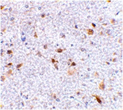
Immunohistsochemistry Analysis: A representative lot detected PD-L2 expression pattern in frozen human tonsil, placenta, fetal thymus and cardiac tissue sections (Brown, J.A., et al. (2003). J. Immunol. 170(3):1257-1266).
Immunocytsochemistry Analysis: A representative lot detected an upregulated PD-L2 immunoreactivity in human CD14+ monocyte-derived macrophages (MDMs) following HIV-BaL infection, but not LPS stimulation by fluorescent immunocytochemistry using paraformaldehyde-fixed MDMs (Rodríguez-García, M., et al. (2011). J. Leukoc. Biol. 89(4) 507–515).
Apoptosis & Cancer
Apoptosis - Additional
Quality
Western Blotting Analysis: 0.5 µg/mL of this antibody detected PD-L2 in 10 µg of human thymus tissue lysate.
Target description
Physical form
Storage and Stability
Handling Recommendations: Upon receipt and prior to removing the cap, centrifuge the vial and gently mix the solution. Aliquot into microcentrifuge tubes and store at -20°C. Avoid repeated freeze/thaw cycles, which may damage IgG and affect product performance.
Other Notes
Disclaimer
Not finding the right product?
Try our Product Selector Tool.
Storage Class Code
12 - Non Combustible Liquids
WGK
WGK 2
Flash Point(F)
Not applicable
Flash Point(C)
Not applicable
Certificates of Analysis (COA)
Search for Certificates of Analysis (COA) by entering the products Lot/Batch Number. Lot and Batch Numbers can be found on a product’s label following the words ‘Lot’ or ‘Batch’.
Already Own This Product?
Find documentation for the products that you have recently purchased in the Document Library.
Our team of scientists has experience in all areas of research including Life Science, Material Science, Chemical Synthesis, Chromatography, Analytical and many others.
Contact Technical Service


