Skip to Content
Merck
All Photos(2)

Key Documents

MABS351-I

Sigma-Aldrich

Anti-FAT10 Antibody, clone 4F1

Synonym(s):

Diubiquitin, UBD, Ubiquitin D

Sign Into View Organizational & Contract Pricing


About This Item

UNSPSC Code:
12352203
NACRES:
NA.41
clone:
4F1, monoclonal
application:
ICC
IP
WB
species reactivity:
human
technique(s):
immunocytochemistry: suitable
immunoprecipitation (IP): suitable
western blot: suitable
citations:

biological source

mouse

Quality Level

antibody form

purified antibody

antibody product type

primary antibodies

clone

4F1, monoclonal

mol wt

calculated mol wt 18 kDa
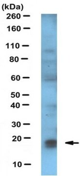
observed mol wt ~18 kDa

purified by

using protein G

species reactivity

human

packaging

antibody small pack of 100

technique(s)

immunocytochemistry: suitable
immunoprecipitation (IP): suitable
western blot: suitable

isotype

IgG2cκ

epitope sequence

Unknown

Protein ID accession no.

UniProt accession no.

storage temp.

2-8°C

Gene Information

human ... UBD(10537)

Specificity

Clone 4F1 is a mouse monoclonal antibody that detects FAT10/Ubiquitin D.

Immunogen

GST-tagged recombinant human FAT10/Ubiquitin D.

Application

Quality Control Testing

Evaluated by Western Blotting in lysate from HepG2 cells treated with IFN and TNF .

Western Blotting Analysis: A 1:1,000 dilution of this antibody detected FAT10 in lysate from HepG2 cells treated with IFN and TNF (200 and 400 units, respectively for 16 h), but not in lysate from untreated HepG2 cells.

Tested Applications

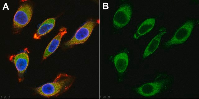
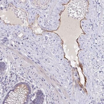
Immunocytochemistry Analysis: A 1:50 dilution from a representative lot detected FAT10/Ubiquitin D in HepG2 cells.

Western Blotting Analysis: A representative lot detected FAT10/Ubiquitin D in Western Blotting application (Aichem, A., et al. (2012). J Cell Sci. 125(Pt19):4576-85; Aichem, A., et al. (2010). Nat Commun.;1:13).

Immunocytochemistry Analysis: A representative lot detected FAT10/Ubiquitin D in Immunocytochemistry applications (Aichem, A., et al. (2012). J Cell Sci. 125(Pt19):4576-85).

Immunoprecipitation Analysis: A representative lot immunoprecipitated FAT10/Ubiquitin D in Immunoprcipitation applications (Aichem, A., et al. (2012). J Cell Sci. 125(Pt19):4576-85).

Note: Actual optimal working dilutions must be determined by end user as specimens, and experimental conditions may vary with the end user.

Target description

Ubiquitin D (UniProt: O15205; also known as Diubiquitin, UBD, Ubiquitin-like protein FAT10) is encoded by the UBD (also known as FAT10) gene (Gene ID: 10537) in human. FAT10 is a ubiquitin-like modifier protein that is involved in various cellular processes, including apoptosis, cell cycle control, and NF- B activation. It is constitutively expressed in mature dendritic cells and B-cells. Its expression is observed in the reticuloendothelial system, the gastrointestinal system, kidney, lung, and prostate gland. It is detected in the nucleus as well as cytoplasm. It has two ubiquitin-like domains (aa 6-81 and 90-163). It can covalently attach to target protein and lead to their degradation by the 26S proteasome, in a NUB1-dependent manner. Its conjugation ability activated by UBA6. It regulates TNF -induced and LPS-mediated activation of NF- B by promoting TNF - mediated proteasomal degradation of I B . It is reported to mediate mitotic non-disjunction and chromosome instability, in long-term in vitro culture and cancers, by abbreviating mitotic phase and impairing the kinetochore localization of mitotic arrest deficient 2 like 1 (MAD2L1) during the prometaphase stage. It mediates apoptosis in a caspase-dependent manner, especially in renal epithelium and tubular cells during renal diseases such as polycystic kidney disease and HIV-associated nephropathy. p62 protein that is involved in autophagy is covalently mono-FAT10ylated at several lysine residues, which leads to proteasomal degradation of p62. Prolonged induction of endogenous FAT10 by pro-inflammatory cytokines leads to a reduction in endogenous p62 levels. Clone 4F1 displays high-affinity for FAT10 and can immunoprecipitate monomeric FAT10 and FAT10-conjugated substrates. (Ref.: Aichem, A., et al. (2012). J. Cell Sci. 125(19); 4576-4585; Ren, J., et al. (2006). J. Biol. Chem. 281(16); 11413-11421; Ross, MJ., et al. (2006). J. Am. Soc. Nephrol. 17(4); 996-1004; Hipp, MS., et al. (2005). Mol. Cell Biol. 25(9); 3483-3491).

Physical form

Purified mouse monoclonal antibody IgG2c in buffer containing 0.1 M Tris-Glycine (pH 7.4), 150 mM NaCl with 0.05% sodium azide.

Reconstitution

1.0 mg/mL. Please refer to guidance on suggested starting dilutions and/or titers per application and sample type.

Storage and Stability

Recommended storage: +2°C to +8°C.

Other Notes

Concentration: Please refer to the Certificate of Analysis for the lot-specific concentration.

Disclaimer

Unless otherwise stated in our catalog or other company documentation accompanying the product(s), our products are intended for research use only and are not to be used for any other purpose, which includes but is not limited to, unauthorized commercial uses, in vitro diagnostic uses, ex vivo or in vivo therapeutic uses or any type of consumption or application to humans or animals.

Not finding the right product?  

Try our Product Selector Tool.

Storage Class Code

12 - Non Combustible Liquids

WGK

WGK 1

Flash Point(F)

Not applicable

Flash Point(C)

Not applicable


Certificates of Analysis (COA)

Search for Certificates of Analysis (COA) by entering the products Lot/Batch Number. Lot and Batch Numbers can be found on a product’s label following the words ‘Lot’ or ‘Batch’.

Already Own This Product?

Find documentation for the products that you have recently purchased in the Document Library.

Visit the Document Library

Our team of scientists has experience in all areas of research including Life Science, Material Science, Chemical Synthesis, Chromatography, Analytical and many others.

Contact Technical Service