Skip to Content
Merck
All Photos(1)

Documents

MAB3073

Sigma-Aldrich

Anti-G Protein Goα Antibody, clone 2A

clone 2A, Chemicon®, from mouse

Synonym(s):

Anti-DEE17, Anti-G-ALPHA-o, Anti-GNAO, Anti-HG1G, Anti-HLA-DQB1, Anti-NEDIM

Sign Into View Organizational & Contract Pricing


About This Item

UNSPSC Code:
12352203
eCl@ss:
32160702
NACRES:
NA.41

biological source

mouse

Quality Level

antibody form

purified immunoglobulin

antibody product type

primary antibodies

clone

2A, monoclonal

species reactivity

rat, bovine, human, guinea pig, mouse

manufacturer/tradename

Chemicon®

technique(s)

western blot: suitable

isotype

IgG1

NCBI accession no.

UniProt accession no.

shipped in

wet ice

target post-translational modification

unmodified

Gene Information

human ... GNAO1(2775)

General description

The previously assigned protein identifier P29777 has been merged into P09471. Full details can be found on the UniProt database.

Specificity

Go-alpha. Strong reactivity with bovine (100%), human (70%), rat (61%) and mouse (54%). Slight reactivity with guinea pig (27%). Specificity confirmed by Western blot versus bovine G-alpha-t, rat Go-alpha, rat Gi-alpha-1, Gi-alpha-2, rat Gi-alpha-3, bovine Gs-alpha, and murine Go-alpha-A and Go-alpha-B. The carboxy-terminal splice variants of Go-alpha (Go-alpha-A and Go-alpha-B) expressed in bacterial lysates were compared for reactivity and found to be equal . As Go-alpha-A and Go-alpha-B have >95% homology to rat Go-alpha-1 and human Go-alpha-2, MAB3073 will detect these G-proteins also.

Immunogen

Bovine brain Go-alpha purified by the method of Sternweis and Robishaw (J. Biol. Chem. [1984] 259, 13806).

Application

Anti-G Protein Goα Antibody, clone 2A is an antibody against G Protein Goα for use in WB.
Research Category
Signaling
Research Sub Category
GPCR, cAMP/cGMP & Calcium Signaling
Western blot 1:3000.

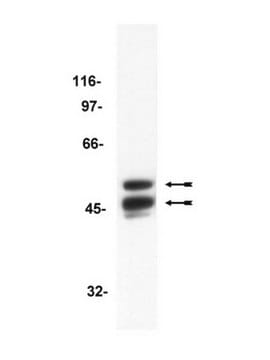
Labels a single protein band of 39 - 42 kDa in bovine and rat membranes.

Optimal working dilutions must be determined by end user.

Physical form

Format: Purified
Purified Immunoglobulin. Liquid in 0.02M PBS, 0.25M NaCl, pH 7.6, containing 0.1% sodium azide as a preservative.

Storage and Stability

Maintain at 2-8°C in undiluted aliquots for upt to 6 months after date of receipt.

During shipment, small volumes of antibody will occasionally become entrapped in the seal of the product vial. For antibodies with volumes of 200 μl or less, we recommend gently tapping the vial on a hard surface or briefly centrifuging the vial in a tabletop centrifuge to dislodge any liquid in the container′s cap.

Other Notes

Concentration: Please refer to the Certificate of Analysis for the lot-specific concentration.

Legal Information

CHEMICON is a registered trademark of Merck KGaA, Darmstadt, Germany

Disclaimer

Unless otherwise stated in our catalog or other company documentation accompanying the product(s), our products are intended for research use only and are not to be used for any other purpose, which includes but is not limited to, unauthorized commercial uses, in vitro diagnostic uses, ex vivo or in vivo therapeutic uses or any type of consumption or application to humans or animals.

Not finding the right product?  

Try our Product Selector Tool.

Storage Class Code

10 - Combustible liquids

WGK

WGK 2

Flash Point(F)

Not applicable

Flash Point(C)

Not applicable


Certificates of Analysis (COA)

Search for Certificates of Analysis (COA) by entering the products Lot/Batch Number. Lot and Batch Numbers can be found on a product’s label following the words ‘Lot’ or ‘Batch’.

Already Own This Product?

Find documentation for the products that you have recently purchased in the Document Library.

Visit the Document Library

Michiel van Wyk et al.
Frontiers in neuroscience, 11, 161-161 (2017-04-21)
Recent studies have demonstrated the safety and efficacy of ocular gene therapy based on adeno-associated viral vectors (AAVs). Accordingly, a surge in promising new gene therapies is entering clinical trials, including the first optogenetic therapy for vision restoration. To date
Zaid Mammo et al.
Investigative ophthalmology & visual science, 56(9), 5074-5086 (2015-08-04)
To demonstrate the utility of speckle variance optical coherence tomography (svOCT), a noninvasive angiographic technique, for evaluating the foveal vasculature. Twelve normal human eyes were imaged with svOCT (1060-nm, 100-kHz custom-built system) and fluorescein angiography (FA; Topcon TRC-50DX with 5.0
Melina A Agosto et al.
eNeuro, 5(3) (2018-07-22)
The chemical signal of light onset, a decrease in glutamate release from rod and cone photoreceptors, is processed by a postsynaptic G protein signaling cascade in ON-bipolar cells (BPCs). The metabotropic glutamate receptor mGluR6, along with other cascade elements, is
Lucien Gazi et al.
British journal of pharmacology, 138(5), 775-786 (2003-03-19)
(1) The human dopamine D(2long) (D(2L)) receptor was expressed with four different G proteins in Sf9 cells using the baculovirus expression system. When co-expressed with G(i)/G(o) G proteins (G(i1)alpha, G(i2)alpha, G(i3)alpha, or G(o)alpha, plus Gbeta(1) and Ggamma(2)), the receptor displayed
Antoine Chaffiol et al.
Current biology : CB, 27(17), 2600-2609 (2017-08-29)
Cone bipolar cells are interneurons that receive synaptic input from cone photoreceptor cells and provide the output of the first synaptic layer of the retina. These cells exhibit center-surround receptive fields, a prototype of lateral inhibition between neighboring sensory cells

Our team of scientists has experience in all areas of research including Life Science, Material Science, Chemical Synthesis, Chromatography, Analytical and many others.

Contact Technical Service