Skip to Content
Merck
All Photos(3)

Key Documents

MAB16985

Sigma-Aldrich

Anti-MCAM Antibody, clone P1H12

clone P1H12, Chemicon®, from mouse

Synonym(s):

CD146 antigen, Cell surface glycoprotein P1H12, Melanoma-associated antigen A32, Melanoma-associated antigen MUC18, S-endo 1 endothelial-associated antigen, melanoma adhesion molecule, melanoma cell adhesion molecule

Sign Into View Organizational & Contract Pricing


About This Item

UNSPSC Code:
12352203
eCl@ss:
32160702
NACRES:
NA.41

biological source

mouse

Quality Level

antibody form

purified immunoglobulin

antibody product type

primary antibodies

clone

P1H12, monoclonal

species reactivity

mouse, canine, human

should not react with

rat

packaging

antibody small pack of 25 μg

manufacturer/tradename

Chemicon®

technique(s)

ELISA: suitable
flow cytometry: suitable
immunocytochemistry: suitable
immunohistochemistry: suitable
immunoprecipitation (IP): suitable
western blot: suitable

isotype

IgG1

suitability

not suitable for immunohistochemistry (Paraffin)

NCBI accession no.

UniProt accession no.

shipped in

ambient

storage temp.

2-8°C

target post-translational modification

unmodified

Gene Information

human ... MCAM(4162)

Related Categories

General description

MUC18 antigen (CD146), a member of the immuoglobulin superfamily, is an intrinsic membrane glycoprotein of 110-120 kDa found on the surface of endothelial cells, bone marrow fibroblasts and various melanomas. MUC18 has been used as a marker of tumor progression in human melanoma because expression in those tumors correlates strongly with poor prognosis and the development of metastic disease. In addition, a number of human T, B and myeloid leukemic cell lines seem to express MUC18. The close structural relationship with N-CAM and related molecules suggests that MUC18 may be also a developmentally regulated cell adhesion molecule (Melanoma adhesion molecule or MCAM).

Specificity

Detects circulating endothelial cells in human. Expected to cross-react with dog & mouse.

Immunogen

Immunization of mice with HUVEC.

Application

Anti-MCAM Antibody, clone P1H12 detects level of MCAM & has been published & validated for use in ELISA, FC, IC, IH, IP & WB.
Immunocytochemistry:
1-10 μg/mL of a previous lot worked in immunocytochemistry.Works best on EDTA or Trypsin lifted endothelial cells.

Immunohistochemistry:
1-10 µg/mL. 4% PFA for 30min RT or <2hrs at 4°C. Block w/ 1%BSA/0.2% tween20/PBS for 30min. Works well in frozen tissue; fixed or unfixed.

Immunoprecipitation:
1-10 μg/mL of a previous lot worked in immunoprecipitation.

ELISA:
1-10 μg/mL of a previous lot worked in ELISA.

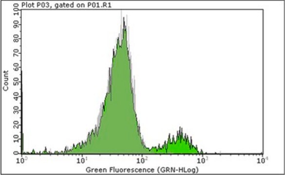
FACS Analysis:
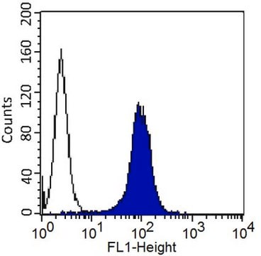
1-10 μg/mL of a previous lot worked in FACS.

Optimal working dilutions must be determined by end user.

Quality

Routinely evaluated by Western Blot on HUVEC lysates.

Western Blot Analysis:
1:500 dilution of this lot detected MCAM on 10μg of HUVEC lysates.

Target description

110 kDa

Physical form

Format: Purified
Purified mouse monoclonal IgG1 in buffer containing 0.02 M PB with 0.25 M NaCl, pH=7.6, with 0.1% sodium azide.

Analysis Note

Control
HUVEC cells.

Other Notes

Concentration: Please refer to the Certificate of Analysis for the lot-specific concentration.

Legal Information

CHEMICON is a registered trademark of Merck KGaA, Darmstadt, Germany

Not finding the right product?  

Try our Product Selector Tool.

Storage Class Code

12 - Non Combustible Liquids

WGK

WGK 2

Flash Point(F)

Not applicable

Flash Point(C)

Not applicable


Certificates of Analysis (COA)

Search for Certificates of Analysis (COA) by entering the products Lot/Batch Number. Lot and Batch Numbers can be found on a product’s label following the words ‘Lot’ or ‘Batch’.

Already Own This Product?

Find documentation for the products that you have recently purchased in the Document Library.

Visit the Document Library

Feng Li et al.
Bone, 47(3), 546-555 (2010-06-24)
Currently, there are conflicting data in literature regarding contribution of bone marrow stromal cells (BMSCs) to bone formation when the cells are systemically delivered in recipient animals. To understand if BMSCs contribute to bone cell phenotype and bone formation in
Yong Wook Park et al.
The American journal of pathology, 165(6), 2087-2098 (2004-12-08)
Thrombospondin 2 (TSP2), a matricellular protein with a primary role in modulating cell-matrix interactions, has been implicated in tissue repair and foreign body responses. Here we show that TSP2 has regulatory function in the chronic inflammatory lesions of rheumatoid arthritis.
Keiich Hirono et al.
Journal of the American College of Cardiology, 48(6), 1257-1264 (2006-09-19)
This study investigated patients with acute Kawasaki disease (KD) to validate myeloid-related protein (MRP)-8/MRP-14 as a marker of disease activity and severity of coronary artery lesion development. Both MRP-8 and -14, which are S100-proteins secreted by activated neutrophils and monocytes
Ivan Depasquale et al.
Cancer cell international, 6, 9-9 (2006-04-01)
Lovastatin and other statins may reduce the development of melanomas. The effects on melanoma cells and their ability to enhance angiogenesis in a co-culture system presented an opportunity to assess whether Lovastatin act on melanoma cells, HUVEC or both types
David M Neskey et al.
Journal of experimental & clinical cancer research : CR, 27, 61-61 (2008-11-06)
Endostatin and anastellin, fragments of collagen type XVIII and fibronectin, respectively, belong to a family of endogenous inhibitors of angiogenesis which inhibit tumor growth and metastasis in a number of mouse models of human cancer. The mechanism of action of

Our team of scientists has experience in all areas of research including Life Science, Material Science, Chemical Synthesis, Chromatography, Analytical and many others.

Contact Technical Service