MABF954
Anti-LAG3 Antibody, clone 4-10-C9
clone 4-10-C9, from mouse
Sinónimos:
Lymphocyte activation gene 3 protein, CD223, LAG-3
About This Item
Productos recomendados
origen biológico
mouse
Nivel de calidad
forma del anticuerpo
purified immunoglobulin
tipo de anticuerpo
primary antibodies
clon
4-10-C9, monoclonal
reactividad de especies
mouse
técnicas
flow cytometry: suitable
immunocytochemistry: suitable
isotipo
IgG2aκ
Nº de acceso NCBI
Nº de acceso UniProt
Condiciones de envío
wet ice
modificación del objetivo postraduccional
unmodified
Información sobre el gen
mouse ... Lag3(16768)
Descripción general
Especificidad
Inmunógeno
Aplicación
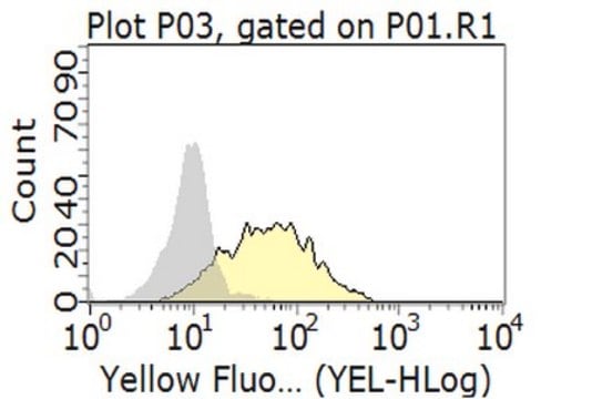
Flow Cytometry Analysis: A representative lot was fluorescently conjugated and detected an increased number of LAG-3-positive cells within the CD4+ and CD8+ populations of infiltrating lymphocytes (TILs) in tumors developed in mice exografted with murine B16 melanoma, MC38 colon adenocarcinoma, or Sa1N fibrosarcoma cells (Woo, S.R., et al. (2012). Cancer Res. 72(4): 917–927).
Flow Cytometry Analysis: A representative lot, pre-conjugated with Alexa Fluor™ 647, detected both surface and intracellular LAG-3 by immunofluorescent staining of non-permeabilized and permeabilized primary murine CD4+ T cells activated in vitro via CD3 & CD28 cross-linking by immobilized antibodies. Pronase treatment of cells prior to permeabilization abolished cell surface staining (Woo, S.R., et al. (2010). Eur. J. Immunol. 40(6):1768-1777).
Flow Cytometry Analysis: A representative lot, pre-conjugated with Alexa Fluor 647, detected a time-dependent recovery of cell surface LAG-3 immunoreactivity on activated murine CD4+ T cells after initial surface LAG-3 degradation by pronase treatment. Protein synthesis inhibitor cycloheximide (Cat. No. 239764) or protein transport inhibitor Brefeldin A (Cat. No. 203729) treatment partially blocked the recovery (Woo, S.R., et al. (2010). Eur. J. Immunol. 40(6):1768-1777).
Immunocytochemistry Analysis: A representative lot detected both surface and intracellular LAG-3 by fluorescent immunocytochemistry staining of non-permeabilized and permeabilized primary murine CD4+ T cells activated in vitro via CD3 & CD28 cross-linking by immobilized antibodies. Pronase treatment of cells prior to permeabilization abolished cell surface staining (Woo, S.R., et al. (2010). Eur. J. Immunol. 40(6):1768-1777).
Immunocytochemistry Analysis: A representative lot detected intracellular LAG-3 immunoreactivity co-localized with those of the early and recycling endosome marker EEA1, as well as endosomal markers Rab11b and Rab27a by fluorescent immunocytochemistry staining of activated murine CD4+ T cells following pronase treatment and permeabilization (Woo, S.R., et al. (2010). Eur. J. Immunol. 40(6):1768-1777).
Calidad
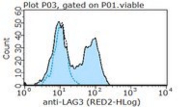
Flow Cytometry Analysis: 1 µg/mL of this antibody detected an induction of LAG-3-positive population in isolated mouse splenocytes following a 3-day 2 µg/mL Concanavalin A (Con A) stimulation.
Descripción de destino
Forma física
Información legal
Not finding the right product?
Try our Herramienta de selección de productos.
Código de clase de almacenamiento
12 - Non Combustible Liquids
Clase de riesgo para el agua (WGK)
WGK 1
Certificados de análisis (COA)
Busque Certificados de análisis (COA) introduciendo el número de lote del producto. Los números de lote se encuentran en la etiqueta del producto después de las palabras «Lot» o «Batch»
¿Ya tiene este producto?
Encuentre la documentación para los productos que ha comprado recientemente en la Biblioteca de documentos.
Nuestro equipo de científicos tiene experiencia en todas las áreas de investigación: Ciencias de la vida, Ciencia de los materiales, Síntesis química, Cromatografía, Analítica y muchas otras.
Póngase en contacto con el Servicio técnico


