Saltar al contenido
Merck
Todas las fotos(1)

Documentos

MABF28

Sigma-Aldrich

Anti-PAR-3 Antibody, clone 8E8

clone 8E8, from mouse

Sinónimos:

Proteinase-activated receptor 3, Coagulation factor II receptor-like 2, Thrombin receptor-like 2

Iniciar sesiónpara Ver la Fijación de precios por contrato y de la organización


About This Item

Código UNSPSC:
12352203
eCl@ss:
32160702
NACRES:
NA.41

origen biológico

mouse

Nivel de calidad

forma del anticuerpo

purified antibody

tipo de anticuerpo

primary antibodies

clon

8E8, monoclonal

reactividad de especies

mouse

reactividad de especies (predicha por homología)

human (based on 100% sequence homology)

técnicas

activity assay: suitable
flow cytometry: suitable

isotipo

IgG2bκ

Nº de acceso NCBI

Nº de acceso UniProt

Condiciones de envío

wet ice

modificación del objetivo postraduccional

unmodified

Información sobre el gen

human ... PARD3(56288)

Descripción general

PAR-3 (also known as Coagulation factor II receptor-like 2, Thrombin receptor-like 2 or F2RL2) is an adapter protein involved in asymmetrical cell division and cell polarization processes. It seems to play a central role in the formation of epithelial tight junctions. Its association with PARD6B may prevent the interaction of PAR-3 with F11R/JAM1, thereby preventing tight junction assembly. The PARD6-PAR-3 complex links GTP bound Rho small GTPases to atypical protein kinase C proteins. PAR-3 interacts with PARD6A and PARD6B. Isoform 2, but not at least isoform 3 interacts with PRKCZ. PAR-3 interacts with PRCKI. PAR-3 forms part of a complex with PARD6A or PARD6B, PRKCI or PRKCZ and CDC42 or RAC1. PAR-3 interacts with F11R/JAM1. Antibodies against PAR-3 are present in sera from patients with cutaneous T cell lymphomas.

Inmunógeno

KLH-conjugated linear peptide corresponding to human PAR-3.

Aplicación

Anti-PAR-3 Antibody, clone 8E8 detects level of PAR-3 & has been published & validated for use in FC, EA.
Flow Cytometry Analysis: A previous lot was used by an independent laboratory in FC. (Petrova, Y., et al. (2008). Centr Eur J Immunol. 33 (1):14-18.)

Activity Assay Analysis (platelet aggregation): A previous lot was used by an independent laboratory in platelet aggregation assay. (Petrova, Y., et al. (2008). Centr Eur J Immunol. 33 (1):14-18.)
Research Category
Infectious Diseases
Research Sub Category
Inflammation & Autoimmune Mechanisms

Calidad

Evaluated by Flow Cytometry in mouse platelets from washed whole blood (heparinized).

Flow Cytometry Analysis: 2 µg of this antibody detected PAR-3 in mouse platelets from washed whole blood (heparinized).

Descripción de destino

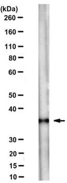
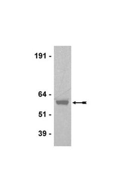
40 kDa calculated

Ligadura / enlace

Replaces: MABS174

Forma física

Format: Purified
Protein G Purified
Purified mouse monoclonal IgG2bκ in buffer containing 0.1 M Tris-Glycine (pH 7.4), 150 mM NaCl with 0.05% sodium azide.

Almacenamiento y estabilidad

Stable for 1 year at 2-8°C from date of receipt.

Nota de análisis

Control
Mouse platelets from washed whole blood (heparinized)

Otras notas

Concentration: Please refer to the Certificate of Analysis for the lot-specific concentration.

Cláusula de descargo de responsabilidad

Unless otherwise stated in our catalog or other company documentation accompanying the product(s), our products are intended for research use only and are not to be used for any other purpose, which includes but is not limited to, unauthorized commercial uses, in vitro diagnostic uses, ex vivo or in vivo therapeutic uses or any type of consumption or application to humans or animals.

¿No encuentra el producto adecuado?  

Pruebe nuestro Herramienta de selección de productos.

Código de clase de almacenamiento

12 - Non Combustible Liquids

Clase de riesgo para el agua (WGK)

WGK 1

Punto de inflamabilidad (°F)

Not applicable

Punto de inflamabilidad (°C)

Not applicable


Certificados de análisis (COA)

Busque Certificados de análisis (COA) introduciendo el número de lote del producto. Los números de lote se encuentran en la etiqueta del producto después de las palabras «Lot» o «Batch»

¿Ya tiene este producto?

Encuentre la documentación para los productos que ha comprado recientemente en la Biblioteca de documentos.

Visite la Librería de documentos

Hiroyuki Nakajima et al.
Journal of cell science, 123(Pt 4), 555-566 (2010-01-28)
Cell-shape change in epithelial structures is fundamental to animal morphogenesis. Recent studies identified myosin-II as the major generator of driving forces for cell-shape changes during morphogenesis. Lulu (Epb41l5) is a major regulator of morphogenesis, although the downstream molecular and cellular
Virginia Fernández et al.
The EMBO journal, 39(21), e105479-e105479 (2020-09-29)
Structural integrity and cellular homeostasis of the embryonic stem cell niche are critical for normal tissue development. In the telencephalic neuroepithelium, this is controlled in part by cell adhesion molecules and regulators of progenitor cell lineage, but the specific orchestration
Michael L Drummond et al.
The Journal of cell biology, 217(9), 3255-3266 (2018-06-28)
Primary cilia are polarized organelles that allow detection of extracellular signals such as Hedgehog (Hh). How the cytoskeleton supporting the cilium generates and maintains a structure that finely tunes cellular response remains unclear. Here, we find that regulation of actin
Kaviya Chinnappa et al.
Science advances, 8(2), eabj4010-eabj4010 (2022-01-13)
The evolutionary expansion and folding of the mammalian cerebral cortex resulted from amplification of progenitor cells during embryonic development. This process was reversed in the rodent lineage after splitting from primates, leading to smaller and smooth brains. Genetic mechanisms underlying

Nuestro equipo de científicos tiene experiencia en todas las áreas de investigación: Ciencias de la vida, Ciencia de los materiales, Síntesis química, Cromatografía, Analítica y muchas otras.

Póngase en contacto con el Servicio técnico