Skip to Content
Merck
All Photos(1)

Documents

311R-1

Sigma-Aldrich

PAX-2 (EP235) Rabbit Monoclonal Primary Antibody

clone EP235, unconjugated, Cell Marque

Sign Into View Organizational & Contract Pricing


About This Item

UNSPSC Code:
12352203
NACRES:
NA.41

biological source

rabbit

Quality Level

100
500

conjugate

unconjugated

antibody form

culture supernatant

antibody product type

primary antibodies

clone

EP235, monoclonal

description

(For In Vitro Diagnostic Use in Select Regions (See Chart))

form

buffered aqueous solution

species reactivity

human

packaging

bottle of 1.0 mL predilute (311R-17)
bottle of 7.0 mL predilute (311R-18)
vial of 0.1 mL concentrate (311R-14)
vial of 0.5 mL concentrate (311R-15)
vial of 1.0 mL concentrate (311R-16)

manufacturer/tradename

Cell Marque

technique(s)

immunohistochemistry (formalin-fixed, paraffin-embedded sections): 1:10-1:50

isotype

IgG

shipped in

wet ice

storage temp.

2-8°C

Gene Information

human ... PAX2(5076)

General description

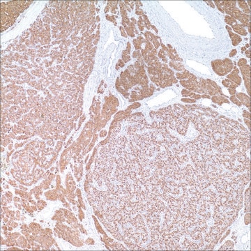
PAX-2 is a homeogene strongly expressed during kidney development. PAX-2 gene is expressed in the metanephric mesenchyma after ureter bud induction and is a key factor for the mesenchyma-epithelium conversion. Animals transgenic for PAX-2 have severe renal abnormalities and cysts but no solid tumoral features. The oncogenic potential of the PAX gene family has been reported <em>in vitro</em> with transformation of cell cultures and <em>in vivo</em> with cell injections in nude mice. Gnarra et al. showed PAX-2 expression in renal carcinoma cell lines and underlined its potential role in cell proliferation in these lines.1 Mazal et al. demonstrated PAX-2 nuclear expression in 88% of clear cell renal cell carcinomas as well as 18% of papillary renal cell carcinoma, and 13% of chromophobe renal cell carcinomas.2 More recently, Chivukula et al. demonstrated utility in distinguishing ovarian serous papillary carcinoma (anti-PAX-2 positive) from breast carcinoma (anti-PAX-2 negative).3

Quality


IVD

IVD

IVD

RUO

Linkage

PAX-2 Positive Control Slides, Product No. 311S, are available for immunohistochemistry (formalin-fixed, paraffin-embedded sections).

Physical form

Solution in Tris Buffer, pH 7.3-7.7, with 1% BSA and <0.1% Sodium Azide.

Preparation Note

Download the IFU specific to your product lot and formatNote: This requires a keycode which can be found on your packaging or product label.

Other Notes

For Technical Service please contact: 800-665-7284 or email: service@cellmarque.com

Legal Information

Cell Marque is a trademark of Merck KGaA, Darmstadt, Germany

Not finding the right product?  

Try our Product Selector Tool.


Certificates of Analysis (COA)

Search for Certificates of Analysis (COA) by entering the products Lot/Batch Number. Lot and Batch Numbers can be found on a product’s label following the words ‘Lot’ or ‘Batch’.

Already Own This Product?

Find documentation for the products that you have recently purchased in the Document Library.

Visit the Document Library

J R Gnarra et al.
Cancer research, 55(18), 4092-4098 (1995-09-15)
Renal cell carcinoma (RCC) is the most common malignancy in the adult kidney. Because RCC is generally thought to arise from the epithelium of the proximal tubules, the expression of Pax-2, a gene required for renal epithelium development, was examined
Mamatha Chivukula et al.
International journal of gynecological pathology : official journal of the International Society of Gynecological Pathologists, 28(6), 570-578 (2009-10-24)
Ovarian serous papillary carcinoma, although rarely metastasizing to the breast, is often challenging based on morphology alone, particularly from the micropapillary variant of breast carcinoma. Gross cystic disease fluid protein-15, although a specific marker, can be negative in up to
Peter R Mazal et al.
Modern pathology : an official journal of the United States and Canadian Academy of Pathology, Inc, 18(4), 535-540 (2004-10-27)
Diagnostic use of antibodies against aquaporin water channel proteins and PAX-2, a nuclear transcription factor in renal development, was tested in 202 renal neoplasms, using tissue microarray technique. Immunohistochemistry for aquaporin-1, aquaporin-2, PAX-2, CD10, and cytokeratin 7 was performed on

Related Content

Diagnostic immunohistochemistry overview highlights its importance in cancer diagnosis and detecting infectious diseases in modern clinical pathology.

Trusted IVDR-ready supplier of high-quality Cell Marque™ antibodies for diagnostic pathology and IHC, ensuring reliability and expert support.

Our team of scientists has experience in all areas of research including Life Science, Material Science, Chemical Synthesis, Chromatography, Analytical and many others.

Contact Technical Service