Skip to Content
Merck
All Photos(1)

Documents

MABT12

Sigma-Aldrich

Anti-Heparan Sulfate Proteoglycan (Perlecan) Antibody, clone 5D7-2E4

clone 5D7-2E4, from mouse

Synonym(s):

heparan sulfate proteoglycan 2 (domain V region), perlecan proteoglycan, basement membrane-specific heparan sulfate proteoglycan core protein, HSPG, PLC

Sign Into View Organizational & Contract Pricing


About This Item

UNSPSC Code:
12352203
eCl@ss:
32160702
NACRES:
NA.41

biological source

mouse

Quality Level

antibody form

purified immunoglobulin

antibody product type

primary antibodies

clone

5D7-2E4, monoclonal

species reactivity

human

technique(s)

immunocytochemistry: suitable
immunohistochemistry: suitable
western blot: suitable

isotype

IgG1κ

NCBI accession no.

UniProt accession no.

shipped in

wet ice

target post-translational modification

unmodified

Gene Information

human ... HSPG2(3339)

General description

Perlecan is one of the extracellular matrix forms of heparan sulfate proteoglycans (HSPGs). It is a large multidomain and binds to and cross-links many extracellular matrix (ECM) components and cell-surface molecules. Perlecan is synthesized by vascular endothelial and smooth muscle cells and deposited in the extracellular matrix within all mammalian tissues. HSPGs differ in their protein cores that are thought to determine the location of HSPG in either the cell membrane (syndecan and glypican) or the extracellular matrix (perlecan and dystroglycan). HSPGs found in the brain are dystroglycan, N-syndecan glypican, and perlecan. The composition of HS isolated from brain differs significantly from that of other organs and is developmentally regulated. Thus, differences in protein cores, disaccharide composition, the extent and position of sulfation, and the number of GAG chains diversify chemical structure and functions of HSPGs. The postulated functions of HSPG include cell proliferation, differentiation, adhesion, migration, and morphogenesis.

Immunogen

Full length recombinant protein corresponding to human Perlecan.

Application

Anti-Heparan Sulfate Proteoglycan (Perlecan) Antibody, clone 5D7-2E4 detects level of Heparan Sulfate Proteoglycan (Perlecan) & has been published & validated for use in IH.
Immunocytochemistry Analysis: A previous lot was used by an independent laboratory in IC. (courtesy of Whitelock, J. Graduate School of Biomedical Engineering, University of New South Wales, Sydney, Australia)

Western Blot Analysis: A previous lot was used by an independent laboratory on a 3-8% Tris-acetate gel in which the band appears above the 460 kDa marker. (courtesy of Whitelock, J. Graduate School of Biomedical Engineering, University of New South Wales, Sydney, Australia)
Research Category
Cell Structure
Research Sub Category
ECM Proteins

Quality

Evaluated by Immunohistochemistry in fresh-frozen normal human tonsil tissue.

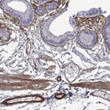
Immunohistochemistry Analysis: 1:300 dilution of the antibody detected Perlecan in fresh-frozen normal human tonsil tissue.

Target description

469 kDa calculated

Physical form

Format: Purified
Protein G
Purified mouse monoclonal IgG1κ in buffer containing 0.1 M Tris-Glycine (pH 7.4), 150 mM NaCl with 0.05% sodium azide.

Storage and Stability

Stable for 1 year at 2-8°C from date of receipt.

Analysis Note

Control
Fresh-frozen normal human tonsil tissue

Other Notes

Concentration: Please refer to the Certificate of Analysis for the lot-specific concentration.

Disclaimer

Unless otherwise stated in our catalog or other company documentation accompanying the product(s), our products are intended for research use only and are not to be used for any other purpose, which includes but is not limited to, unauthorized commercial uses, in vitro diagnostic uses, ex vivo or in vivo therapeutic uses or any type of consumption or application to humans or animals.

Not finding the right product?  

Try our Product Selector Tool.

Storage Class Code

12 - Non Combustible Liquids

WGK

WGK 1

Flash Point(F)

Not applicable

Flash Point(C)

Not applicable


Certificates of Analysis (COA)

Search for Certificates of Analysis (COA) by entering the products Lot/Batch Number. Lot and Batch Numbers can be found on a product’s label following the words ‘Lot’ or ‘Batch’.

Already Own This Product?

Find documentation for the products that you have recently purchased in the Document Library.

Visit the Document Library

Sokratis A Apostolidis et al.
Frontiers in immunology, 9, 2191-2191 (2018-10-18)
Objective: The mechanisms that lead to endothelial cell (EC) injury and propagate the vasculopathy in Systemic Sclerosis (SSc) are not well understood. Using single cell RNA sequencing (scRNA-seq), our goal was to identify EC markers and signature pathways associated with
Veronica De Paolis et al.
International journal of molecular sciences, 23(14) (2022-07-28)
The cellular heterogeneity of the tumor environment of breast cancer (BC) is extremely complex and includes different actors such as neoplastic, stromal, and immunosuppressive cells, which contribute to the chemical and mechanical modification of the environment surrounding the tumor-exasperating immune-escaping
Shoichiro Maeda et al.
British journal of pharmacology, 179(20), 4857-4877 (2022-07-08)
Chondroitin sulfate proteoglycan (CSPG) constitutes the neurogenic niche in the hippocampus. The reduction of hippocampal neurogenesis is involved in ageing-related cognitive decline and dementia. The purpose of this study is to find candidates that improve cognitive function by analysing the
Sofiane Hamidi et al.
Development (Cambridge, England), 147(3) (2020-02-06)
The pluripotent epiblast gives rise to all tissues and organs in the adult body. Its differentiation starts at gastrulation, when the epiblast generates mesoderm and endoderm germ layers through epithelial-mesenchymal transition (EMT). Although gastrulation EMT coincides with loss of epiblast
Steffen Rickelt et al.
Matrix biology : journal of the International Society for Matrix Biology, 71-72, 10-27 (2018-05-08)
The diversity of extracellular matrix (ECM) proteins encoded in mammalian genomes and detected by proteomic analyses generates a need for well validated antibodies against these proteins. We present characterization of a large number of antibodies against ECM proteins, from both

Our team of scientists has experience in all areas of research including Life Science, Material Science, Chemical Synthesis, Chromatography, Analytical and many others.

Contact Technical Service