Skip to Content
Merck
All Photos(3)

Key Documents

HPA008855

Sigma-Aldrich

Anti-MUC1 antibody produced in rabbit

enhanced validation

Prestige Antibodies® Powered by Atlas Antibodies, affinity isolated antibody, buffered aqueous glycerol solution

Synonym(s):

Anti-ADMCKD, Anti-ADMCKD1, Anti-CD227, Anti-MCD, Anti-MCKD, Anti-MCKD1, Anti-PEM, Anti-PUM

Sign Into View Organizational & Contract Pricing


About This Item

MDL number:
UNSPSC Code:
12352203
Human Protein Atlas Number:
NACRES:
NA.43

biological source

rabbit

Quality Level

conjugate

unconjugated

antibody form

affinity isolated antibody

antibody product type

primary antibodies

clone

polyclonal

product line

Prestige Antibodies® Powered by Atlas Antibodies

form

buffered aqueous glycerol solution

species reactivity

human

enhanced validation

orthogonal RNAseq
Learn more about Antibody Enhanced Validation

technique(s)

immunohistochemistry: 1:50- 1:200

UniProt accession no.

application(s)

research pathology

shipped in

wet ice

storage temp.

−20°C

target post-translational modification

unmodified

Gene Information

human ... MUC1(4582)

Immunogen

mucin 1 isoform 5 precursor recombinant protein epitope signature tag (PrEST)

Sequence
AVCQCRRKNYGQLDIFPARDTYHPMSEYPTYHTHGRYVPPSSTDRSPYEKVSAGNGGSSLSYTNPAVAATSANL

Application

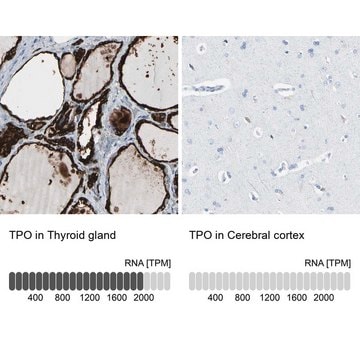
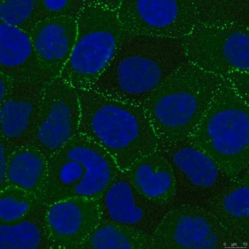
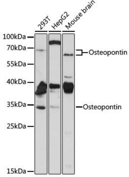
Anti-MUC1 antibody produced in rabbit, a Prestige Antibody, is developed and validated by the Human Protein Atlas (HPA) project . Each antibody is tested by immunohistochemistry against hundreds of normal and disease tissues. These images can be viewed on the Human Protein Atlas (HPA) site by clicking on the Image Gallery link. The antibodies are also tested using immunofluorescence and western blotting. To view these protocols and other useful information about Prestige Antibodies and the HPA, visit sigma.com/prestige.

Biochem/physiol Actions

MUC1 (mucin 1) is localized to the chromosomal locus which is the third most frequently rearranged site in non-Hodgkin′s lymphoma (NHL). Chromosomal translocation results in the activation of this gene, which is responsible for tumorigenesis in lymphomas. Due to chromosomal rearrangements, this gene is deregulated in leukemias, sarcomas, and epithelial tumors. This gene leads to the switching of SMAD3 signaling from tumor suppressive (pSmad3C/p21(WAF1)) to oncogenic pSmad3L/c-Myc. This leads to the activation of c-Jun NH2-terminal kinase (JNK) signaling in hepatocellular carcinoma (HCC). In cutaneous T-cell lymphoma (CTCL), this protein maintains redox balance, which prevents ROS (reactive oxygen species)-mediated apoptosis. Thus, it has potential as a therapeutic target in the same.

Features and Benefits

Prestige Antibodies® are highly characterized and extensively validated antibodies with the added benefit of all available characterization data for each target being accessible via the Human Protein Atlas portal linked just below the product name at the top of this page. The uniqueness and low cross-reactivity of the Prestige Antibodies® to other proteins are due to a thorough selection of antigen regions, affinity purification, and stringent selection. Prestige antigen controls are available for every corresponding Prestige Antibody and can be found in the linkage section.

Every Prestige Antibody is tested in the following ways:
  • IHC tissue array of 44 normal human tissues and 20 of the most common cancer type tissues.
  • Protein array of 364 human recombinant protein fragments.

Linkage

Corresponding Antigen APREST70726

Physical form

Solution in phosphate-buffered saline, pH 7.2, containing 40% glycerol and 0.02% sodium azide

Legal Information

Prestige Antibodies is a registered trademark of Merck KGaA, Darmstadt, Germany

Disclaimer

Unless otherwise stated in our catalog or other company documentation accompanying the product(s), our products are intended for research use only and are not to be used for any other purpose, which includes but is not limited to, unauthorized commercial uses, in vitro diagnostic uses, ex vivo or in vivo therapeutic uses or any type of consumption or application to humans or animals.

Not finding the right product?  

Try our Product Selector Tool.

Storage Class Code

10 - Combustible liquids

WGK

WGK 1

Flash Point(F)

Not applicable

Flash Point(C)

Not applicable

Personal Protective Equipment

dust mask type N95 (US), Eyeshields, Gloves

Certificates of Analysis (COA)

Search for Certificates of Analysis (COA) by entering the products Lot/Batch Number. Lot and Batch Numbers can be found on a product’s label following the words ‘Lot’ or ‘Batch’.

Already Own This Product?

Find documentation for the products that you have recently purchased in the Document Library.

Visit the Document Library

V G Dyomin et al.
Blood, 95(8), 2666-2671 (2001-02-07)
The band 1q21 is among the most common sites affected by chromosomal translocations in lymphoid, myeloid, epithelial, and sarcomatous lesions. In non-Hodgkin's lymphoma (NHL), translocations and duplications affecting this chromosomal site are frequently, but not exclusively, seen in association with
Devon A Lawson et al.
Nature, 526(7571), 131-135 (2015-09-30)
Despite major advances in understanding the molecular and genetic basis of cancer, metastasis remains the cause of >90% of cancer-related mortality. Understanding metastasis initiation and progression is critical to developing new therapeutic strategies to treat and prevent metastatic disease. Prevailing
Qiongshu Li et al.
Oncotarget, 6(6), 4253-4265 (2015-02-26)
Mucin1 (MUC1) is a transmembrane glycoprotein that acts as an oncogene in human hepatic tumorigenesis. Hepatocellular carcinoma (HCC) cells often gain advantage by reducing the tumor-suppressive activity of transforming growth factor beta (TGF-β) together with stimulation of its oncogenic activity
Salvia Jain et al.
Blood, 126(3), 354-362 (2015-06-07)
Cutaneous T-cell lymphoma (CTCL) is an aggressive neoplasm with limited treatments for patients with advanced disease. The mucin 1 C-terminal subunit (MUC1-C) oncoprotein plays a critical role in regulating cell proliferation, apoptosis, and protection from cytotoxic injury mediated by reactive
Chien-Ting Wu et al.
Cell, 186(1), 112-130 (2022-12-30)
How SARS-CoV-2 penetrates the airway barrier of mucus and periciliary mucins to infect nasal epithelium remains unclear. Using primary nasal epithelial organoid cultures, we found that the virus attaches to motile cilia via the ACE2 receptor. SARS-CoV-2 traverses the mucus

Our team of scientists has experience in all areas of research including Life Science, Material Science, Chemical Synthesis, Chromatography, Analytical and many others.

Contact Technical Service