Skip to Content
Merck
All Photos(3)

Key Documents

E1031

Sigma-Aldrich

Anti-ERGIC-53/p58 antibody produced in rabbit

affinity isolated antibody, buffered aqueous solution

Synonym(s):

Anti-ERGIC53, Anti-F5F8D, Anti-FMFD1, Anti-LMAN1, Anti-MCFD1, Anti-MR60, Anti-gp58

Sign Into View Organizational & Contract Pricing


About This Item

MDL number:
UNSPSC Code:
12352203
NACRES:
NA.41

biological source

rabbit

Quality Level

conjugate

unconjugated

antibody form

affinity isolated antibody

antibody product type

primary antibodies

clone

polyclonal

form

buffered aqueous solution

mol wt

antigen ~58 kDa

species reactivity

mouse, rat, human

technique(s)

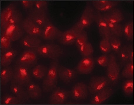
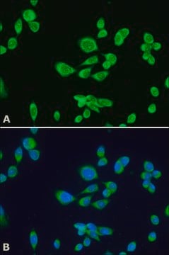
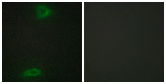
indirect immunofluorescence: 5-10 μg/mL using human HepG2 or rat NRK cells
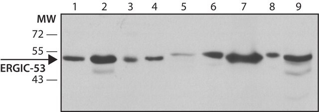
western blot (chemiluminescent): 0.5-1.0 μg/mL using whole cell extract of mouse NIH3T3 cells

UniProt accession no.

shipped in

dry ice

storage temp.

−20°C

target post-translational modification

unmodified

Gene Information

human ... LMAN1(3998)
mouse ... Lman1(70361)
rat ... Lman1(116666)

General description

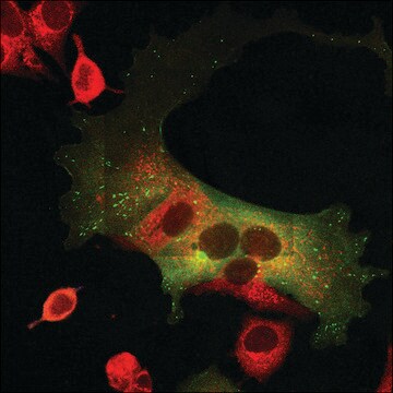
ERGIC (ER-Golgi intermediate compartment) is related to type I membrane marker protein ERGIC-53 and rat homolog is referred as (p58) . ERGIC is a dynamic membrane system made up of tubulo vesicular clusters near to ER exit site which facilitates transport of protein from ER to Golgi. Anti-ERGIC-53/ (p58) antibody can be used as a primary antibody (diluted 1: 125) in fluorescence microscopy. It may also be used for western blotting. Rabbit anti-ERGIC-53/ (p58) antibodies react specifically with ERGIC-53/ (p58) of rat, mouse and human.

Immunogen

synthetic peptide corresponding to amino acids 158-170 of rat p58 with N-terminal added cysteine, conjugated to KLH. The corresponding sequence is identical in mouse and human.

Application

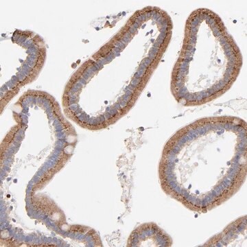
Anti-ERGIC-53/ (p58) antibody can be used in immunoblotting and immunofluorescence.

Physical form

Solution in 0.01 M phosphate buffered saline, pH 7.4, containing 1% bovine serum albumin and 15 mM sodium azide.

Disclaimer

Unless otherwise stated in our catalog or other company documentation accompanying the product(s), our products are intended for research use only and are not to be used for any other purpose, which includes but is not limited to, unauthorized commercial uses, in vitro diagnostic uses, ex vivo or in vivo therapeutic uses or any type of consumption or application to humans or animals.

Not finding the right product?  

Try our Product Selector Tool.

Storage Class Code

10 - Combustible liquids

WGK

nwg

Flash Point(F)

Not applicable

Flash Point(C)

Not applicable


Certificates of Analysis (COA)

Search for Certificates of Analysis (COA) by entering the products Lot/Batch Number. Lot and Batch Numbers can be found on a product’s label following the words ‘Lot’ or ‘Batch’.

Already Own This Product?

Find documentation for the products that you have recently purchased in the Document Library.

Visit the Document Library

A Schweizer et al.
The Journal of cell biology, 107(5), 1643-1653 (1988-11-01)
Purified Golgi membranes of the human intestinal adenocarcinoma cell line Caco-2 were used as an antigen to produce a monoclonal antibody, G1/93, which specifically labels a tubulovesicular compartment near the cis side of the Golgi apparatus, including the first cis-cisterna
Aaron B Bowen et al.
eLife, 6 (2017-09-07)
Neurons face the challenge of regulating the abundance, distribution and repertoire of integral membrane proteins within their immense, architecturally complex dendritic arbors. While the endoplasmic reticulum (ER) supports dendritic translation, most dendrites lack the Golgi apparatus (GA), an essential organelle
AT-1 is the ER membrane acetyl-CoA transporter and is essential for cell viability.
Mary Cabell Jonas, Mariana Pehar
Journal of Cell Science, 23, 3378-3388 (2010)
J Saraste et al.
The Journal of cell biology, 105(5), 2021-2029 (1987-11-01)
A 58-kD cis-Golgi protein has been identified by generating polyclonal antibodies against heavy (cis) Golgi subfractions. Total microsomes isolated from rat pancreatic homogenates were subfractionated to yield a rough microsomal fraction (B1) and three smooth membrane subfractions (B2-B4) enriched in
Adam K Walker et al.
PloS one, 8(11), e81170-e81170 (2013-12-07)
In amyotrophic lateral sclerosis (ALS) and frontotemporal lobar degeneration, TAR DNA binding protein 43 (TDP-43) accumulates in the cytoplasm of affected neurons and glia, where it associates with stress granules (SGs) and forms large inclusions. SGs form in response to

Our team of scientists has experience in all areas of research including Life Science, Material Science, Chemical Synthesis, Chromatography, Analytical and many others.

Contact Technical Service