Skip to Content
Merck
All Photos(5)

Documents

MAB5356

Sigma-Aldrich

Anti-Rhodopsin Antibody, CT, last 9 amino acids, clone Rho 1D4

clone Rho 1D4, Chemicon®, from mouse

Synonym(s):

Anti-CSNBAD1, Anti-OPN2, Anti-RP4

Sign Into View Organizational & Contract Pricing


About This Item

UNSPSC Code:
12352203
eCl@ss:
32160702
NACRES:
NA.41

biological source

mouse

Quality Level

antibody form

purified immunoglobulin

antibody product type

primary antibodies

clone

Rho 1D4, monoclonal

species reactivity

vertebrates

manufacturer/tradename

Chemicon®

technique(s)

immunocytochemistry: suitable
immunohistochemistry: suitable
immunoprecipitation (IP): suitable
western blot: suitable

isotype

IgG1

NCBI accession no.

UniProt accession no.

shipped in

wet ice

target post-translational modification

unmodified

Gene Information

human ... RHO(6010)

Specificity

Recognizes Rhodopsin. Reacts with both native and recombinant protein. No known reactivity to other proteins.

Immunogen

Bovine rhodopsin
Epitope: C-terminus, last 9 amino acids

Application

Anti-Rhodopsin Antibody, C-terminus, last 9 amino acids, clone Rho 1D4 detects level of Rhodopsin & has been published & validated for use in IC, IH, IP & WB.
Research Category
Neuroscience
Research Sub Category
Sensory & PNS
Western blot: 1:100-1:500 using ECL. Suggested dilution buffer is PBS and blocking buffer is PBS containing 5% milk. Recognizes 36 kDa monomers, dimmers and trimers depending on sample preparation. Boiling induces the aggregation of rhodopsin.

Western blot of isolated bovine rod outer segment labeled for rhodopsin (~85% of rod outer segment protein) with the rho 1D4 antibody. Amount of rod outer segments applied to the SDS gel: (a) 2.5 mg; (b) 0.63 mg; (c) 0.16 mg; (d) 0.04 mg. At higher protein quantities, dimers, trimers and tetramers of rhodopsin can be observed along with the monomer.

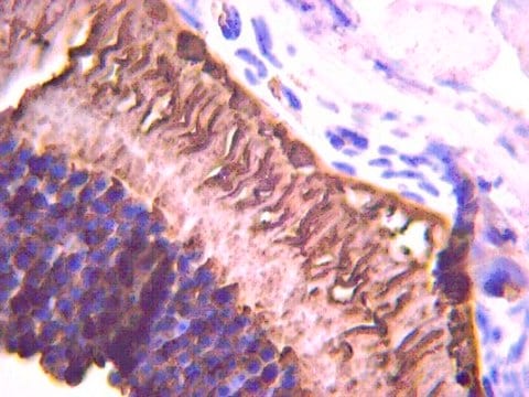
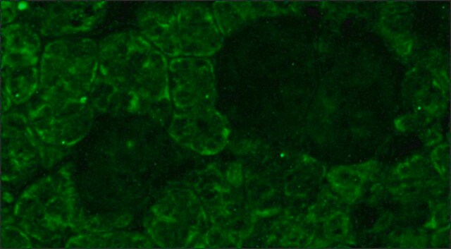
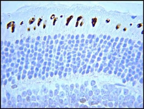
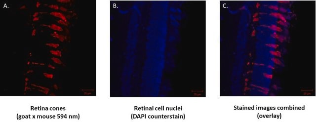
Immunohisto/cytochemistry: 1:100-1:500 on paraformaldehyde and glutaraldehyde fixed frozen tissue sections. Preferred fixation is paraformaldehyde for 4 hours at 2-8°C. Suggested permeabilization method is 0.2% Triton X-100.

Immunoprecipitation

Optimal working dilutions must be determined by the end user.

Target description

48 kDa

Physical form

Format: Purified
Protein A Purified mouse immunoglobulin in 20 mM sodium phosphate, 250 mM NaCl, pH. 7.6, with 0.1% sodium azide as a preservative.
Protein A purified

Storage and Stability

Maintain for 1 year at 2–8°C from date of shipment. Aliquot to avoid repeated freezing and thawing. For maximum recovery of product, centrifuge the original vial after thawing and prior to removing the cap.

Analysis Note

Control
Eye

Other Notes

Concentration: Please refer to the Certificate of Analysis for the lot-specific concentration.

Legal Information

CHEMICON is a registered trademark of Merck KGaA, Darmstadt, Germany

Disclaimer

Unless otherwise stated in our catalog or other company documentation accompanying the product(s), our products are intended for research use only and are not to be used for any other purpose, which includes but is not limited to, unauthorized commercial uses, in vitro diagnostic uses, ex vivo or in vivo therapeutic uses or any type of consumption or application to humans or animals.

Not finding the right product?  

Try our Product Selector Tool.

Storage Class Code

10 - Combustible liquids

WGK

WGK 2

Flash Point(F)

Not applicable

Flash Point(C)

Not applicable


Certificates of Analysis (COA)

Search for Certificates of Analysis (COA) by entering the products Lot/Batch Number. Lot and Batch Numbers can be found on a product’s label following the words ‘Lot’ or ‘Batch’.

Already Own This Product?

Find documentation for the products that you have recently purchased in the Document Library.

Visit the Document Library

Tomomi Sakai et al.
Journal of autoimmunity, 81, 13-23 (2017-03-23)
TREX1/DNASE III, the most abundant 3'-5' DNA exonuclease in mammalian cells, is tail-anchored on the endoplasmic reticulum (ER). Mutations at the N-terminus affecting TREX1 DNase activity are associated with autoimmune and inflammatory conditions such as Aicardi-Goutières syndrome (AGS). Mutations in
M Keith Wyatt et al.
PloS one, 8(6), e68088-e68088 (2013-07-11)
Age-related macular degeneration (AMD) is a major cause of vision loss. It is associated with development of characteristic plaque-like deposits (soft drusen) in Bruch's membrane basal to the retinal pigment epithelium (RPE). A sequence variant (Y402H) in short consensus repeat
Bernice H Wong et al.
The Journal of biological chemistry, 291(20), 10501-10514 (2016-03-25)
Eye photoreceptor membrane discs in outer rod segments are highly enriched in the visual pigment rhodopsin and the ω-3 fatty acid docosahexaenoic acid (DHA). The eye acquires DHA from blood, but transporters for DHA uptake across the blood-retinal barrier or
Clémence Bernard et al.
Human molecular genetics, 23(7), 1742-1753 (2013-11-16)
In the human, mutations of OTX2 (Orthodenticle homeobox 2 transcription factor) translate into eye malformations of variable expressivity (even between the two eyes of the same individual) and incomplete penetrance, suggesting the existence of subtle thresholds in OTX2 activity. We
M Corton et al.
Scientific reports, 6, 35370-35370 (2016-10-14)
Retinitis pigmentosa (RP), the most frequent form of inherited retinal dystrophy is characterized by progressive photoreceptor degeneration. Many genes have been implicated in RP development, but several others remain to be identified. Using a combination of homozygosity mapping, whole-exome and

Our team of scientists has experience in all areas of research including Life Science, Material Science, Chemical Synthesis, Chromatography, Analytical and many others.

Contact Technical Service