indirect immunofluorescence: 5-10 μg/mL using differentiated mouse NIH3T3-L1 cells western blot (chemiluminescent): 2.5-5 μg/mL using whole extract of differentiated mouse NIH3T3-L1 cells
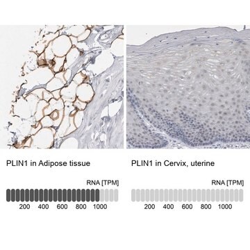
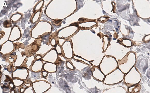
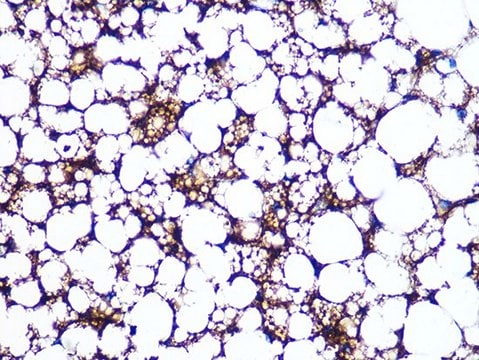
Perilipin is an intracellular neutral lipid storage droplet surface protein in white and brown fat adipocytes. It is also found in lower quantity coating droplets in steroidogenic-cells of the adrenal cortex, ovaries, and testicular Leydig cells, on the surface of smaller droplets containing cholesteryl esters.
Specificity
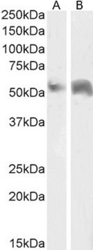
Rabbit polyclonal anti-Perilipin A/B antibody recognizes mouse Perilipin A/B by immunoblotting (46 kDa and ~62 kDa) and indirect immunofluorescence. Detection of the perilipin A and B bands by immunoblotting is specifically inhibited with the immunizing peptide.
Immunogen
synthetic peptide corresponding amino acid residues 6-17 of mouse and rat perilipin A/B with C-terminal added cysteine, conjugated to KLH. The corresponding human sequence differs by two residues.
Application
Anti-Perilipin A/B antibody produced in rabbit has been used in immunoblotting and immunofluorescence staining.[1]
Rabbit polyclonal anti-Perilipin A/B antibody is used to tag Perilipin A/B for detection and quantitation by immunocytochemical and immunohistochemical (IHC) techniques. It is used as a probe to determine the presence and roles of Perilipin A/B in the regulation of triacylglycerol storage in adipocytes.
Biochem/physiol Actions
Perilipin is a gatekeeper protein that is involved in regulating triacylglycerol storage in adipocyte through the suppression of basal lipolysis apparently through protecting triacylglycerol against hydrolysis. Perilipin also enhances cAMP-dependent protein kinase (PKA)-stimulated lipolysis by hormone-sensitive lipase (HSL) and non-HSLs. Perilipin knockout mice exhibit reduced adipose tissue mass and resistance to diet induced obesity. Their lipid storage droplets are coated with adipose differentiation-related protein (ADRP, adipophilin), that is devoid of phosphorylation by PKA.
Physical form
Solution in 0.01 M phosphate buffered saline, pH 7.4, containing 15 mM sodium azide.
Disclaimer
Unless otherwise stated in our catalog or other company documentation accompanying the product(s), our products are intended for research use only and are not to be used for any other purpose, which includes but is not limited to, unauthorized commercial uses, in vitro diagnostic uses, ex vivo or in vivo therapeutic uses or any type of consumption or application to humans or animals.
The American journal of pathology, 187(12), 2627-2634 (2017-09-19)
Fatty degeneration of skeletal muscle leads to muscle weakness and loss of function. Preventing fatty degeneration in skeletal muscle is important, but no drug has been used clinically. In this study, we performed drug repositioning using human platelet-derived growth factor
During the aging process, bone marrow mesenchymal stem cells (BMSCs) exhibit declined osteogenesis accompanied by excess adipogenesis, which will lead to osteoporosis. Here, we report that the H3 lysine 36 trimethylation (H3K36me3), catalyzed by histone methyltransferase SET-domain-containing 2 (SETD2), regulates
The amino and carboxyl termini of perilipin a facilitate the storage of triacylglycerols
Garcia A, et al.
The Journal of Biological Chemistry, 279(9), 8409-8416 (2004)
Reestablishment of energy balance in a male mouse model with POMC neuron deletion of BMPR1A
MyoD and Myf5 are fundamental regulators of skeletal muscle lineage determination in the embryo, and their expression is induced in satellite cells following muscle injury. MyoD and Myf5 are also expressed by satellite cell precursors developmentally, although the relative contribution
Our team of scientists has experience in all areas of research including Life Science, Material Science, Chemical Synthesis, Chromatography, Analytical and many others.