AC-0368A
p62 (Sequestosome-1) (EP396) Rabbit Monoclonal Primary Antibody
Sign Into View Organizational & Contract Pricing
All Photos(1)
About This Item
UNSPSC Code:
12352203
NACRES:
NA.41
Recommended Products
biological source
rabbit
Quality Level
antibody form
Ig fraction of antiserum
clone
monoclonal
description
For In Vitro Diagnostic Use in Select Regions (See Chart)
form
buffered aqueous solution
technique(s)
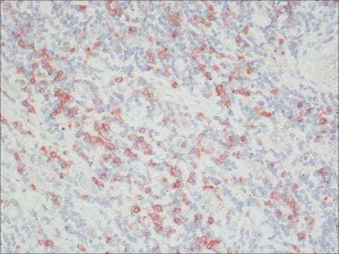
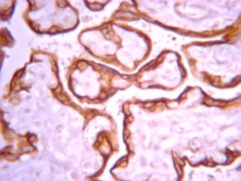
immunohistochemistry (formalin-fixed, paraffin-embedded sections): 1:100-1:200
shipped in
wet ice
storage temp.
2-8°C
visualization
(nuclear,cytoplasm)
General description
P62, also known as Sequestosome-1, is a ubiquitin-binding adapter protein involved in the delivery of ubiquitin-bound protein and organelles to the autophagosome for lysosomal degradation during autophagy. Mutations in p62 have been linked to Paget′s disease and neurodegenerative disorders. P62 is typically degraded during the autophagy process, but can accumulate and/or overexpressed in autophagy-deficient cells. Presence of p62 cytosolic aggregate is used as a marker for autophagy deficiency. Defective autophagy increase oxidative stress and may play a role in tumorigenesis. P62 immunohistochemistry (IHC) diagnostic utility was established for identifying hepatocellular carcinoma (HCC). P62 demonstrates superior sensitivity for HCC compared against glypican-3. Positive p62 staining was found in 100% of HCC but were negative in all non-tumor areas and cirrhotic nodules. Furthermore, a panel consisting of p62, aminoacylase 1, and glypican-3 provides high sensitivity (93.8%) and specificity (95.2%) in the differential diagnosis between well differentiated HCC and high grade dysplastic nodules. P62 expression can also aid in diagnosing drug-induced autophagic vacuolar myopathies. An optimal threshold of at least 11.5% positive fibers provided 100% sensitivity and 100% specificity for autophagic myopathy. While current diagnostic criteria require identification of autophagic vacuoles by electron microscopy, diagnosis with p62 IHC could expedite the process and reduce costliness and large sampling variance associated with electron microscopy.
Quality
 IVD |  IVD |  IVD |  RUO |
Physical form
Solution in Tris Buffer, pH 7.3-7.7, with 1% BSA and <0.1% Sodium Azide
Other Notes
For a copy of the IFU and CofA contact IVDorder@ABCAM.com
For Technical Service please contact: 800-665-7284 or email: service@cellmarque.com
For Technical Service please contact: 800-665-7284 or email: service@cellmarque.com
Storage Class Code
10 - Combustible liquids
WGK
WGK 2
Flash Point(F)
Not applicable
Flash Point(C)
Not applicable
Choose from one of the most recent versions:
Certificates of Analysis (COA)
Lot/Batch Number
Sorry, we don't have COAs for this product available online at this time.
If you need assistance, please contact Customer Support.
Already Own This Product?
Find documentation for the products that you have recently purchased in the Document Library.
Our team of scientists has experience in all areas of research including Life Science, Material Science, Chemical Synthesis, Chromatography, Analytical and many others.
Contact Technical Service