
C1801
Monoclonal Anti-Cytokeratin, pan antibody produced in mouse
ascites fluid, clone PCK-26
Sinónimos:
Monoclonal Anti-pan-Cytokeratin
About This Item
Productos recomendados


origen biológico
mouse
Nivel de calidad
conjugado
unconjugated
forma del anticuerpo
ascites fluid
tipo de anticuerpo
primary antibodies
clon
PCK-26, monoclonal
contiene
15 mM sodium azide
reactividad de especies
human, chicken, snake, hamster, pig, goat, feline, bovine, carp, rat, rabbit, canine, lizard, sheep, mouse, guinea pig
técnicas
immunohistochemistry (formalin-fixed, paraffin-embedded sections): 1:300
immunohistochemistry (frozen sections): suitable
indirect immunofluorescence: 1:300 using protease-idgested, formalin-fixed, paraffin-embedded sections of human or animal tissues
western blot: suitable
isotipo
IgG1
Condiciones de envío
dry ice
temp. de almacenamiento
−20°C
modificación del objetivo postraduccional
unmodified
Información sobre el gen
bovine ... Krt1(100301161)
dog ... Krt1(444857)
human ... KRT1(3848) , KRT1(3848) , KRT1(3848) , KRT5(3852) , KRT5(3852) , KRT5(3852) , KRT6A(3868) , KRT6A(3868) , KRT6A(3868) , KRT6B(3854) , KRT6B(3854) , KRT6B(3854) , KRT8(3856) , KRT8(3856) , KRT8(3856)
mouse ... KRT1(16678) , KRT1(16678) , KRT1(16678) , Krt1(16678) , Krt5(110308) , Krt5(110308) , Krt5(110308) , Krt6a(16687) , Krt6a(16687) , Krt6a(16687) , Krt6b(16688) , Krt6b(16688) , Krt6b(16688) , Krt8(16691) , Krt8(16691) , Krt8(16691)
rat ... Krt1(300250) , Krt2-5(369017) , Krt2-5(369017) , Krt2-5(369017) , Krt2-8(25626) , Krt2-8(25626) , Krt2-8(25626)
¿Está buscando productos similares? Visita Guía de comparación de productos
Descripción general
Especificidad
Inmunógeno
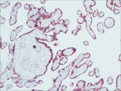
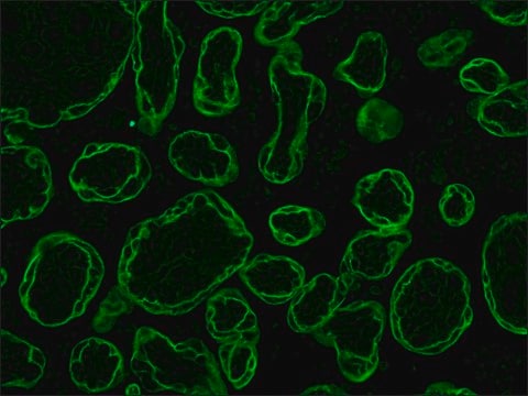
Aplicación
Monoclonal Anti-Cytokeratin, pan may be used for the localization of cytokeratins using various immunochemical assays such as immunoblotting, dot blotting, and immunohistochemstry (immunofluorescence and immunoenzymatic staining).
A minimum antibody titer of 1:300 was determined by indirect immunofluorescent staining of protease digested, formalin-fixed, paraffin-embedded sections of human or animal tissues.
Acciones bioquímicas o fisiológicas
Cláusula de descargo de responsabilidad
¿No encuentra el producto adecuado?
Pruebe nuestro Herramienta de selección de productos.
Opcional
Producto relacionado
Código de clase de almacenamiento
10 - Combustible liquids
Clase de riesgo para el agua (WGK)
WGK 3
Punto de inflamabilidad (°F)
Not applicable
Punto de inflamabilidad (°C)
Not applicable
Elija entre una de las versiones más recientes:
Certificados de análisis (COA)
¿No ve la versión correcta?
Si necesita una versión concreta, puede buscar un certificado específico por el número de lote.
¿Ya tiene este producto?
Encuentre la documentación para los productos que ha comprado recientemente en la Biblioteca de documentos.
Los clientes también vieron
Nuestro equipo de científicos tiene experiencia en todas las áreas de investigación: Ciencias de la vida, Ciencia de los materiales, Síntesis química, Cromatografía, Analítica y muchas otras.
Póngase en contacto con el Servicio técnico