Saltar al contenido
Merck
Todas las fotos(1)

Documentos

FCMAB205F

Sigma-Aldrich

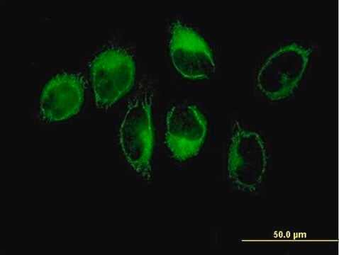
Milli-Mark® Anti-CD68-FITC Antibody, clone KP1

clone KP1, Milli-Mark®, from mouse

Sinónimos:

Macrosialin, Gp110

Iniciar sesiónpara Ver la Fijación de precios por contrato y de la organización


About This Item

UNSPSC Code:
12352203
eCl@ss:
32160702
NACRES:
NA.44

biological source

mouse

Quality Level

conjugate

FITC conjugate

antibody form

purified immunoglobulin

antibody product type

primary antibodies

clone

KP1, monoclonal

species reactivity

human

manufacturer/tradename

Milli-Mark®

technique(s)

flow cytometry: suitable

isotype

IgG1κ

NCBI accession no.

UniProt accession no.

shipped in

wet ice

target post-translational modification

unmodified

Gene Information

human ... CD68(968)

General description

CD68 is highly glycosylated lysosomal membrane protein with an Mr of 110,000. The CD68 protein belongs to a family of lysosomal glycoprotein (LGP)/plasma membrane shuttling proteins that play a role in endocytosis and/or lysosomal trafficking. CD68 is expressed strongly in cytoplasmic granules, and weakly on the surface of macrophages, monocytes, neutrophils, basophils, and NK cells. CD68 can also be found in the cytoplasm of non-hematopoietic tissues, especially the liver, and renal glomeruli, and tubules (Goyert, 1997).

Specificity

Antibody recognizes Human CD68 antigen.

Immunogen

Lysosomal fraction of human lung macrophages

Application

Research Category
Inflammation & Immunology
Research Sub Category
Immunoglobulins & Immunology
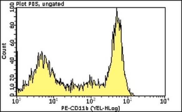
This Milli-Mark Anti-CD68-FITC Antibody, clone KP1 is validated for use in FC for the detection of CD68.

Quality

Evaluated by flow cytometry using human lymphocytes

Target description

110 kDa (Goyert, 1997)

Physical form

Protein A purified
Purified Mouse monoclonal IgG1 conjugated to FITC in 0.05M Tris-HCl buffer with 1% BSA, Sodium Azide at 0.097% at pH 7.2.

Storage and Stability

Maintain refrigerated at 2-8°C protected from light in undiluted aliquots for up to 6 months from date of receipt.

Analysis Note

Control
Human Lymphocytes

Legal Information

MILLI-MARK is a registered trademark of Merck KGaA, Darmstadt, Germany

Disclaimer

Unless otherwise stated in our catalog or other company documentation accompanying the product(s), our products are intended for research use only and are not to be used for any other purpose, which includes but is not limited to, unauthorized commercial uses, in vitro diagnostic uses, ex vivo or in vivo therapeutic uses or any type of consumption or application to humans or animals.

¿No encuentra el producto adecuado?  

Pruebe nuestro Herramienta de selección de productos.

Storage Class

12 - Non Combustible Liquids

wgk_germany

WGK 2

flash_point_f

Not applicable

flash_point_c

Not applicable


Certificados de análisis (COA)

Busque Certificados de análisis (COA) introduciendo el número de lote del producto. Los números de lote se encuentran en la etiqueta del producto después de las palabras «Lot» o «Batch»

¿Ya tiene este producto?

Encuentre la documentación para los productos que ha comprado recientemente en la Biblioteca de documentos.

Visite la Librería de documentos

Manuel Moreno-Valladares et al.
Aging, 12(19), 18928-18941 (2020-10-14)
The presence and functional role of T cell infiltration in human brain parenchyma with normal aging and neurodegeneration is still under intense debate. Recently, CD8+ cells have been shown to infiltrate the subventricular zone in humans and mice with a
Marion Duriez et al.
Journal of clinical and translational hepatology, 8(4), 359-370 (2021-01-16)
Background and Aims: To better understand nonalcoholic steatohepatitis (NASH) disease progression and to evaluate drug targets and compound activity, we undertook the development of an in vitro 3D model to mimic liver architecture and the NASH environment. Methods: We have

Nuestro equipo de científicos tiene experiencia en todas las áreas de investigación: Ciencias de la vida, Ciencia de los materiales, Síntesis química, Cromatografía, Analítica y muchas otras.

Póngase en contacto con el Servicio técnico