Saltar al contenido
Merck
Todas las fotos(3)

Documentos

ABT129

Sigma-Aldrich

Anti-Calponin-1 Antibody

from rabbit, purified by affinity chromatography

Sinónimos:

Calponin-1, Basic calponin, Calponin H1, smooth muscle

Iniciar sesiónpara Ver la Fijación de precios por contrato y de la organización


About This Item

UNSPSC Code:
12352203
eCl@ss:
32160702
NACRES:
NA.41

biological source

rabbit

Quality Level

antibody form

affinity isolated antibody

antibody product type

primary antibodies

clone

polyclonal

purified by

affinity chromatography

species reactivity

human

species reactivity (predicted by homology)

sheep (based on 100% sequence homology), bovine (based on 100% sequence homology), chimpanzee (based on 100% sequence homology)

technique(s)

immunofluorescence: suitable
immunohistochemistry: suitable (paraffin)
western blot: suitable

NCBI accession no.

UniProt accession no.

shipped in

wet ice

target post-translational modification

unmodified

Gene Information

human ... CNN1(1264)

General description

Calponin 1 is a 34 kDa protein that is predominantly found in smooth muscle tissue. It is a high-affinity actin-binding protein, but also interacts with tropomyosin, and calmodulin to modulate muscle contraction. When bound to F-actin, it inhibits the activity of the actin-activated myosin ATPase. Calponin 1 is regulated by reversible phosphorylation involving PKC and CAMKII kinases, and type 2A phosphatases. In non-muscle cells, calponin 1 may play a role in cell migration, motility, and proliferation.

Specificity

Other homologies: Rat (92% sequence homology) and Mouse (83% sequence homology).
This antibody recognizes the C-terminus of Calponin-1.

Immunogen

Epitope: C-terminus
KLH-conjugated linear peptide corresponding to the C-terminus of human Calponin-1.

Application

Immunohistochemistry Analysis: A 1:1,000 dilution from a reprsesentative lot detected Calponin-1 in human seminal vesicle tissue.

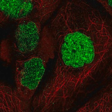
Immunofluorescence Analysis: A 1:500 dilution from a representative lot detected Calponin-1 in smooth muscle cells from human seminal vesicle tissue.
Research Category
Cell Structure
Research Sub Category
Cytoskeleton
This Anti-Calponin-1 Antibody is validated for use in Western Blotting, IHC(P), Immunofluorescence for the detection of Calponin-1.

Quality

Evaluated by Western Blot in human aortic smooth muscle tissue.

Western Blot Analysis: 0.1 µg/mL of this antibody detected Calponin-1 in human aortic smooth muscle tissue.

Target description

~33 kDa observed

Linkage

Replaces: 04-589

Physical form

Affinity purified
Purified rabbit polyclonal in buffer containing 0.1 M Tris-Glycine (pH 7.4), 150 mM NaCl with 0.05% sodium azide.

Storage and Stability

Stable for 1 year at 2-8°C from date of receipt.

Analysis Note

Control
Human aortic smooth muscle tissue

Other Notes

Concentration: Please refer to the Certificate of Analysis for the lot-specific concentration.

Disclaimer

Unless otherwise stated in our catalog or other company documentation accompanying the product(s), our products are intended for research use only and are not to be used for any other purpose, which includes but is not limited to, unauthorized commercial uses, in vitro diagnostic uses, ex vivo or in vivo therapeutic uses or any type of consumption or application to humans or animals.

¿No encuentra el producto adecuado?  

Pruebe nuestro Herramienta de selección de productos.

Optional

Storage Class

12 - Non Combustible Liquids

wgk_germany

WGK 1

flash_point_f

Not applicable

flash_point_c

Not applicable


Certificados de análisis (COA)

Busque Certificados de análisis (COA) introduciendo el número de lote del producto. Los números de lote se encuentran en la etiqueta del producto después de las palabras «Lot» o «Batch»

¿Ya tiene este producto?

Encuentre la documentación para los productos que ha comprado recientemente en la Biblioteca de documentos.

Visite la Librería de documentos

Shunchi Zhang et al.
Molecular medicine (Cambridge, Mass.), 28(1), 121-121 (2022-10-04)
Stent implantation-induced neointima formation is a dominant culprit in coronary artery disease treatment failure after percutaneous coronary intervention. Ferroptosis, an iron-dependent regulated cell death, has been associated with various cardiovascular diseases. However, the effect of ferroptosis on neointima formation remains
Edward P Carter et al.
Breast cancer research : BCR, 19(1), 50-50 (2017-04-22)
3D modelling fulfils a critical role in research, allowing for complex cell behaviour and interactions to be studied in physiomimetic conditions. With tissue banks becoming established for a number of cancers, researchers now have access to primary patient cells, providing
TRNT-1 Deficiency Is Associated with Loss of tRNA Integrity and Imbalance of Distinct Proteins.
Fatica, et al.
Genes, 14 (2023)
Sudip Kumar Paul et al.
Nature communications, 15(1), 4772-4772 (2024-06-11)
The underlying mechanisms of atherosclerosis, the second leading cause of death among Werner syndrome (WS) patients, are not fully understood. Here, we establish an in vitro co-culture system using macrophages (iMφs), vascular endothelial cells (iVECs), and vascular smooth muscle cells

Nuestro equipo de científicos tiene experiencia en todas las áreas de investigación: Ciencias de la vida, Ciencia de los materiales, Síntesis química, Cromatografía, Analítica y muchas otras.

Póngase en contacto con el Servicio técnico