Skip to Content
Merck
All Photos(5)

Documents

H3284

Sigma-Aldrich

Anti-Histone Deacetylase 1 (HDAC1) antibody produced in rabbit

IgG fraction of antiserum, buffered aqueous solution

Synonym(s):

Anti-GON-10, Anti-HD1, Anti-KDAC1, Anti-RPD3, Anti-RPD3L1

Sign Into View Organizational & Contract Pricing


About This Item

MDL number:
UNSPSC Code:
12352203
NACRES:
NA.41

biological source

rabbit

Quality Level

conjugate

unconjugated

antibody form

IgG fraction of antiserum

antibody product type

primary antibodies

clone

polyclonal

form

buffered aqueous solution

mol wt

antigen 65 kDa

species reactivity

human, mouse

packaging

antibody small pack of 25 μL

technique(s)

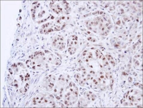
immunohistochemistry (formalin-fixed, paraffin-embedded sections): 1:500 using human lymph node sections
immunoprecipitation (IP): 5-10 μL using whole lysate of NIH3T3 cells
microarray: suitable
western blot: 1:20,000 using nuclear extract from HeLa human epithelioid carcinoma cells

UniProt accession no.

shipped in

dry ice

storage temp.

−20°C

target post-translational modification

unmodified

Gene Information

human ... HDAC1(3065)
mouse ... Hdac1(433759)

General description

Acetylation is a histone modification in which epsilon amino group of conserve lysine residue is modified with enzymes known as histone acetyltransferases (HATs). HATs are present in two forms nuclear and cytoplasmic. Histone deacetylases are competating enzymes of HATs and are often associated with transcriptional repression and nucleosome condensation. Anti-Histone Deacetylase 1 (HDAC1) antibody can be used in chromatin immunoprecipitation. . This antibody can also be used in double immunofloresence labeling. Anti-Histone Deacetylase 1 (HDAC1) antibody specifically recognizes histone deacetylase 1and reacts specifically with HDAC1 of human, rat and mouse.
Histone acetylation is a dynamic process whose levels are determined by the net activities of HATs and the competing enzymes histone deacetylases (HDACs). Both activities are associated with the nuclear matrix. Six or seven different mammalian HDACs have been described. HDAC1, HDAC2 and HDAC3 are similar to yeast Rpd3 protein, while HDAC4, HDAC5 and HDAC6 are similar to yeast Hda1 protein. Histone deacetylases activities were often, but not always, associated with transcriptional repression and nucleosomal condensations. HDAC1, HDAC2 and several other HDACs are the catalytic subunits of different multiprotein regulatory complexes.
Histone deacetylase 1 (HDAC1) is located in the nucleus. It belongs to class I of histone deacetylases. HDAC1 is located on human chromosome 1p35.

Specificity

Anti-Histone Deacetylase 1 specifically recognizes histone deacetylase 1 by immunoblotting and immunoprecipitation (65 kDa). An additional band of lower molecular weight may be detected in some cell line extracts by immunoblotting. Staining of HDAC1 by immunoblotting is specifically inhibited with the immunizing peptide. The antibody is also useful for the detection of HDAC1 by immunohistochemistry. The epitope(s) recognized by the antibody is resistant to routine formalin-fixation and paraffin-embedding, unless there is a protease digestion. The antibody reacts with HDAC1 of human, rat and mouse origin.

Immunogen

synthetic peptide corresponding to amino acids 466-482 of human and mouse HDAC1.

Application

Anti-Histone Deacetylase 1 (HDAC1) antibody can be used in immunoblotting; immunoprecipitation and immunohistochemistry. It is also useful in chromatin immunoprecipitation.
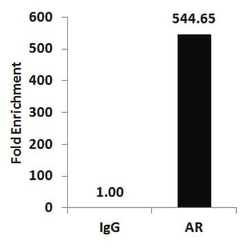
Anti-Histone Deacetylase 1 (HDAC1) antibody has been used in immunohistochemical staining and chromatin immunoprecipitation (ChIP) analyses.
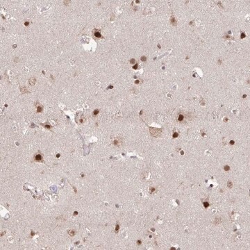
Anti-Histone Deacetylase 1 is suitable for immunoblotting; immunoprecipitation and immunohistochemistry. (IHC)
Immunoblotting: a minimum working dilution of 1:20,000 is determined using a nuclear extract of HeLa human epithelioid carcinoma cell line.
Immunoblotting: a minimum working dilution of 1:2,000 is determined using a whole extract of PC-12 rat pheochromocytoma cell line.
Immunoprecipitation: a recommended working volume of 5-10 ml is determined using a whole lysate of NIH 3T3 cells.
Indirect immunoperoxidase staining: a minimum working dilution of 1:500 is determined using protease-digested, formalin-fixed, paraffin-embedded human lymph node sections.

Biochem/physiol Actions

Histone deacetylase 1 (HDAC1) prevents the infection of influenza A virus (IAV). The protein plays important roles in DNA repair, splicing, regulation of gene expression and cell division. Overexpression of this gene can be seen in cancers like breast, renal cell cancer, and classical Hodgkin′s lymphoma.

Physical form

Solution in 0.01 M phosphate bufffered saline, pH 7.4, containing 15 mM sodium azide.

Disclaimer

Unless otherwise stated in our catalog or other company documentation accompanying the product(s), our products are intended for research use only and are not to be used for any other purpose, which includes but is not limited to, unauthorized commercial uses, in vitro diagnostic uses, ex vivo or in vivo therapeutic uses or any type of consumption or application to humans or animals.

Not finding the right product?  

Try our Product Selector Tool.

Storage Class Code

12 - Non Combustible Liquids

WGK

WGK 2

Flash Point(F)

Not applicable

Flash Point(C)

Not applicable

Personal Protective Equipment

dust mask type N95 (US), Eyeshields, Gloves

Certificates of Analysis (COA)

Search for Certificates of Analysis (COA) by entering the products Lot/Batch Number. Lot and Batch Numbers can be found on a product’s label following the words ‘Lot’ or ‘Batch’.

Already Own This Product?

Find documentation for the products that you have recently purchased in the Document Library.

Visit the Document Library

Histone deacetylase HDAC1 expression correlates with the progression and prognosis of lung cancer: a meta-analysis
Cao L L, et al.
Medicine, 96(31) (2017)
Influenza A virus dysregulates host histone deacetylase 1 that inhibits viral infection in lung epithelial cells
Nagesh P T and Husain M
Journal of virology, JVI-00126 (2016)
Ludovica Lospinoso Severini et al.
Cancers, 11(10) (2019-10-12)
: Pharmacological Hedgehog (Hh) pathway inhibition has emerged as a valuable anticancer strategy. A number of small molecules able to block the pathway at the upstream receptor Smoothened (Smo) or the downstream effector glioma-associated oncogene 1 (Gli1) has been designed
Direct p53 transcriptional repression: in vivo analysis of CCAAT-containing G2/M promoters
Imbriano C, et al.
Molecular and cellular biology, 25(9), 3737-3751 (2005)
Barbara Ulmasov et al.
Cellular and molecular gastroenterology and hepatology, 2(4), 499-518 (2017-02-09)
Pancreatic stellate cells (PSCs) regulate the development of chronic pancreatitis (CP) and are activated by the cytokine transforming growth factor β (TGFB). Integrins of the αv family promote TGFB signaling in mice, probably by interacting with the Arg-Gly-Asp (RGD) sequence

Our team of scientists has experience in all areas of research including Life Science, Material Science, Chemical Synthesis, Chromatography, Analytical and many others.

Contact Technical Service