Skip to Content
Merck
All Photos(1)

Documents

MABC40

Sigma-Aldrich

Anti-LAMP-2 Antibody, clone ABL-93

clone ABL-93, from rat

Synonym(s):

lysosomal-associated membrane protein 2, CD107 antigen-like family member B, CD107b antigen

Sign Into View Organizational & Contract Pricing


About This Item

UNSPSC Code:
12352203
eCl@ss:
32160702
NACRES:
NA.41

biological source

rat

Quality Level

antibody form

purified immunoglobulin

antibody product type

primary antibodies

clone

ABL-93, monoclonal

species reactivity

mouse

technique(s)

immunofluorescence: suitable
immunoprecipitation (IP): suitable
western blot: suitable

isotype

IgG2aκ

NCBI accession no.

UniProt accession no.

shipped in

wet ice

target post-translational modification

unmodified

Gene Information

mouse ... Lamp2(16784)

General description

Lysosome-associated membrane protein 2 (LAMP-2) is a highly glycosolated protein belonging to the lysosome-associated membrane protein family. This group pf proteins composes a significant portion of the lysosomal membrane and are considered crucial mediators for cellular adhesion by interacting with carbohydrate receptors. LAMP-2 is a profuse and critical component of the lysosomal membrane as it has been implicated in playing a role in lysosomal biogenesis. It also appears to be involved in later stages of phagocytosis and autophagy, where it may act to protect the lysosomal membrane from being digested, assist in signal transduction, moderate levels of acidity within the lysosomal environment, and participate in adhesion. Mutations in LAMP-2 expression have been associated with lysosomal storage diseases or LSDs, along with Danon disease, an X-linked disorder characterized by skeletal myopathy, mental retardation, and hypertrophic cardiomyopathy.

Specificity

This antibody recognizes LAMP-2.

Immunogen

Purified glycoproteins from NIH/3T3 cells, corresponding to mouse LAMP-2.

Application

Anti-LAMP-2 Antibody, clone ABL-93 is an antibody against LAMP-2 for use in WB, IP & IF.
Immunoprecipitation Analysis: This antibody has been shown to immunoprecipitate LAMP-2 in HaNIH cells, (Chen, 1985).

Immunofluorescence Analysis: This antibody has been shown to detect LAMP-2 in mouse brain, (Ferguson, 2009).
Research Category
Apoptosis & Cancer
Research Sub Category
Apoptosis - Additional

Quality

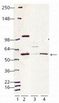
Evaluated by Western Blot in NIH/3T3 cell lysate.

Western Blot Analysis: 1 µg/mL of this antibody detected LAMP-2 in 10 µg of NIH/3T3 cell lysate.

Target description

~ 80 kDa observed MW. The calculated MW is 45 kDa LAMP-2 is heavily glycosoated and has been routinely observed at MW′s of 80-120 kDa based on publication data, (Chen, 1985; Ferguson, 2009).

Physical form

Format: Purified
Protein G
Purified rat monoclonal IgG2aκ in buffer containing 0.1 M Tris-Glycine (pH 7.4), 150 mM NaCl with 0.05% sodium azide.

Storage and Stability

Stable for 1 year at 2-8°C from date of receipt.

Analysis Note

Control
NIH/3T3 cell lysate

Other Notes

Concentration: Please refer to the Certificate of Analysis for the lot-specific concentration.

Disclaimer

Unless otherwise stated in our catalog or other company documentation accompanying the product(s), our products are intended for research use only and are not to be used for any other purpose, which includes but is not limited to, unauthorized commercial uses, in vitro diagnostic uses, ex vivo or in vivo therapeutic uses or any type of consumption or application to humans or animals.

Not finding the right product?  

Try our Product Selector Tool.

Storage Class Code

12 - Non Combustible Liquids

WGK

WGK 1

Flash Point(F)

Not applicable

Flash Point(C)

Not applicable


Certificates of Analysis (COA)

Search for Certificates of Analysis (COA) by entering the products Lot/Batch Number. Lot and Batch Numbers can be found on a product’s label following the words ‘Lot’ or ‘Batch’.

Already Own This Product?

Find documentation for the products that you have recently purchased in the Document Library.

Visit the Document Library

Celia A McKee et al.
Scientific reports, 12(1), 1796-1796 (2022-02-04)
An emerging link between circadian clock function and neurodegeneration has indicated a critical role for the molecular clock in brain health. We previously reported that deletion of the core circadian clock gene Bmal1 abrogates clock function and induces cell-autonomous astrocyte activation. Regulation of
Debashis Dutta et al.
Cell reports, 40(2), 111058-111058 (2022-07-14)
This study underlines the importance of treadmill exercise in reducing α-synuclein (α-syn) spreading in the A53T brain and protecting nigral dopaminergic neurons. Preformed α-syn fibril (PFF) seeding in the internal capsule of young A53T α-syn mice leads to increased spreading
Imene Jaadane et al.
Journal of cellular and molecular medicine, 21(12), 3453-3466 (2017-07-01)
Ageing and alteration of the functions of the retinal pigment epithelium (RPE) are at the origin of lost of vision seen in age-related macular degeneration (AMD). The RPE is known to be vulnerable to high-energy blue light. The white light-emitting
Hui Shen et al.
Neural regeneration research, 18(8), 1770-1776 (2023-02-09)
Recent studies have shown that microglia/macrophages and astrocytes can mediate synaptic phagocytosis through the MER proto-oncokinase in developmental or stroke models, but it is unclear whether the same mechanism is also active in traumatic brain injury. In this study, we
Abhilash P Appu et al.
Journal of inherited metabolic disease, 42(5), 944-954 (2019-04-27)
Mutations in at least 13 different genes (called CLNs) underlie various forms of neuronal ceroid lipofuscinoses (NCLs), a group of the most common neurodegenerative lysosomal storage diseases. While inactivating mutations in the CLN1 gene, encoding palmitoyl-protein thioesterases-1 (PPT1), cause infantile

Our team of scientists has experience in all areas of research including Life Science, Material Science, Chemical Synthesis, Chromatography, Analytical and many others.

Contact Technical Service