Accéder au contenu
Merck
Toutes les photos(1)

Key Documents

M6190

Sigma-Aldrich

Monoclonal Anti-MYOD1 antibody produced in mouse

clone 5.2F, purified immunoglobulin, buffered aqueous solution

Synonyme(s) :

Anti-Myogenic Differentiation Antigen 1

Se connecterpour consulter vos tarifs contractuels et ceux de votre entreprise/organisme


About This Item

Numéro MDL:
Code UNSPSC :
12352203
Nomenclature NACRES :
NA.41

Source biologique

mouse

Niveau de qualité

Conjugué

unconjugated

Forme d'anticorps

purified immunoglobulin

Type de produit anticorps

primary antibodies

Clone

5.2F, monoclonal

Forme

buffered aqueous solution

Poids mol.

antigen 34 kDa

Espèces réactives

human, rat, chicken, mouse

Concentration

1.0 mg/mL

Technique(s)

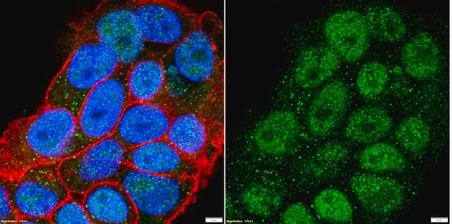
immunocytochemistry: suitable
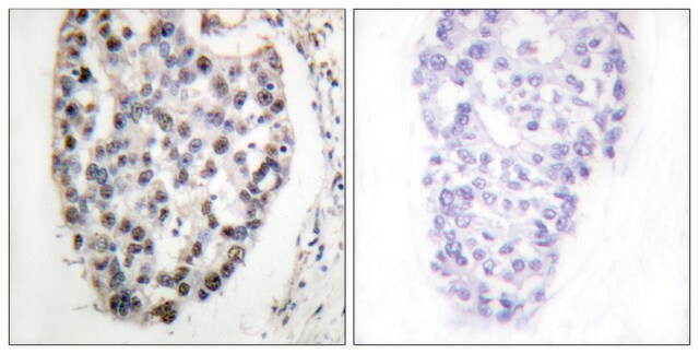
immunohistochemistry (formalin-fixed, paraffin-embedded sections): 2-4 μg/mL
immunohistochemistry (frozen sections): 2-4 μg/mL
immunoprecipitation (IP): 2 μg using 1 mg protein lysate
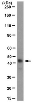
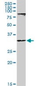
western blot: 1 μg/mL (reacts with the ~45 kDa protein)

Isotype

IgG2a

Numéro d'accès UniProt

Conditions d'expédition

wet ice

Température de stockage

−20°C

Informations sur le gène

human ... MYOD1(4654)
mouse ... Myod1(17927)
rat ... Myod1(337868)

Description générale

Myogenic differentiation antigen 1 (MYOD1) is a nuclear protein which is expressed in skeletal muscles. It is part of the basic helix-loop-helix (bHLH) family of transcription factors. The gene encoding it is localized on human chromosome 11.

Immunogène

recombinant mouse MyoD1 protein.

Application

Monoclonal Anti-MYOD1 antibody produced in mouse has been used in:
  • immunofluorescence staining at a 1:50 dilution
  • western blotting
  • immunostaining at a 1:300 dilution

Actions biochimiques/physiologiques

Myogenic differentiation antigen 1 (MYOD1) maybe involved in recruitment of enzymes like acetyltransferases and methyltransferases to myogenic enhancers in the human genome. It takes part in the regeneration of muscles and mediates muscle cell differentiation by activating cell cycle arrest.

Forme physique

Solution in phosphate buffered saline containing 0.08% sodium azide.

Clause de non-responsabilité

Unless otherwise stated in our catalog or other company documentation accompanying the product(s), our products are intended for research use only and are not to be used for any other purpose, which includes but is not limited to, unauthorized commercial uses, in vitro diagnostic uses, ex vivo or in vivo therapeutic uses or any type of consumption or application to humans or animals.

Vous ne trouvez pas le bon produit ?  

Essayez notre Outil de sélection de produits.

Code de la classe de stockage

10 - Combustible liquids

Classe de danger pour l'eau (WGK)

nwg

Point d'éclair (°F)

Not applicable

Point d'éclair (°C)

Not applicable


Certificats d'analyse (COA)

Recherchez un Certificats d'analyse (COA) en saisissant le numéro de lot du produit. Les numéros de lot figurent sur l'étiquette du produit après les mots "Lot" ou "Batch".

Déjà en possession de ce produit ?

Retrouvez la documentation relative aux produits que vous avez récemment achetés dans la Bibliothèque de documents.

Consulter la Bibliothèque de documents

Shujie Chen et al.
Bio-protocol, 9(14), e3313-e3313 (2019-07-20)
Myofiber isolation followed with ex vivo culture could recapitulate and visualize satellite cells (SCs) activation, proliferation, and differentiation. This approach could be taken to understand the physiology of satellite cells and the molecular mechanism of regulatory factors, in terms of
Bahar Shahidi et al.
JOR spine, 3(2), e1087-e1087 (2020-07-03)
Many chronic musculoskeletal conditions are associated with loss of muscle volume and quality, resulting in functional decline. While atrophy has long been implicated as the mechanism of muscle loss in these conditions, recent evidence has emerged demonstrating a degenerative phenotype
Roy Blum
Journal of cellular biochemistry, 115(11), 1855-1867 (2014-06-07)
The early 1980s revelation of cis-acting genomic elements, known as transcriptional enhancers, is still regarded as one of the fundamental discoveries in the genomic field. However, only with the emergence of genome-wide techniques has the genuine biological scope of enhancers
Bolin Cai et al.
Cellular & molecular biology letters, 29(1), 9-9 (2024-01-05)
Skeletal muscle development is pivotal for animal growth and health. Recently, long noncoding RNAs (lncRNAs) were found to interact with chromatin through diverse roles. However, little is known about how lncRNAs act as chromatin-associated RNAs to regulate skeletal muscle development.
Xiangyu Sui et al.
Frontiers in oncology, 12, 1040112-1040112 (2022-11-18)
Skeletal muscle atrophy is the major hallmark of cancer cachexia. The mechanisms underlying muscle wasting remain elusive in cachectic patients. Our research seeks to identify differentially expressed genes (DEGs) between non-cachectic and cachectic cancer patients and elucidate their functions. We

Notre équipe de scientifiques dispose d'une expérience dans tous les secteurs de la recherche, notamment en sciences de la vie, science des matériaux, synthèse chimique, chromatographie, analyse et dans de nombreux autres domaines..

Contacter notre Service technique