Accéder au contenu
Merck
Toutes les photos(2)

Documents

C2931

Sigma-Aldrich

Monoclonal Anti-Cytokeratin, pan antibody produced in mouse

clone C-11, ascites fluid

Synonyme(s) :

Monoclonal Anti-Pan Cytokeratin

Se connecterpour consulter vos tarifs contractuels et ceux de votre entreprise/organisme


About This Item

Code UNSPSC :
12352203
Nomenclature NACRES :
NA.41

Source biologique

mouse

Niveau de qualité

Conjugué

unconjugated

Forme d'anticorps

ascites fluid

Type de produit anticorps

primary antibodies

Clone

C-11, monoclonal

Contient

15 mM sodium azide

Espèces réactives

bovine, mouse, frog, human, kangaroo rat, rat

Technique(s)

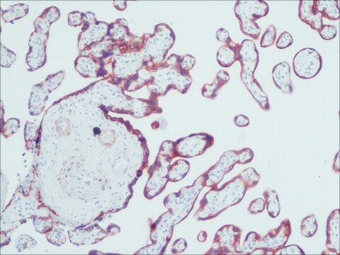
immunohistochemistry (formalin-fixed, paraffin-embedded sections): suitable using protease-digested sections of human or animal tissues
immunohistochemistry (frozen sections): suitable
indirect immunofluorescence: 1:400 using protease-digested, formalin-fixed, paraffin-embedded sections of human or animal tissues
western blot: suitable

Isotype

IgG1

Conditions d'expédition

dry ice

Température de stockage

−20°C

Modification post-traductionnelle de la cible

unmodified

Vous recherchez des produits similaires ? Visite Guide de comparaison des produits

Description générale

Intermediate-sized filaments are abundant cytoplasmic structural proteins in most vertebrate cells. Cytokeratins, a group of at least 29 different proteins, are characteristic of epithelial and trichocytic cells. Cytokeratins 4, 5, 6 and 8 are members of the type II neutral-to-basic subfamily. Cytokeratin peptide 4 (59 kDa) is the secondary type II keratin expressed in noncornified stratified squamous epithelia. Cytokeratin peptide 5 (58 kDa) is the primary type II keratin in stratified epithelia, while cytokeratin type 8 (52 kDa) is a major type II keratin in simple epithelia. Cytokeratin 6 (56 kDa) is a "hyperproliferation" cytokeratin expressed in tissues with natural or pathological high turnover. Cytokeratins 10, 13 and 18 are members of the type I acidic subfamily. Cytokeratin peptide 10 (56 kDa) is the secondary type I keratin expressed in cornified epithelia. Cytokeratin 13 (54 kDa) is the secondary type I keratin expressed in non-cornified stratified squamous epithelia. Cytokeratin 18 (45 kDa) is the primary type I keratin expressed in simple epithelial cells.
Monoclonal Anti-Pan Cytokeratin (clone C-11) recognizes human cytokeratins 4, 5, 6, 8, 10, 13 and 18 in immunoblotting. The antibody reacts with simple, cornifying and non-cornifying squamous epithelia and pseudostratified epithelia. It does not react with non-epithelial normal human tissues. This antibody can be applied to methanol or acetone-fixed, frozen sections, and to formalin-fixed, paraffin-embedded human tissues. Increased staining intensity is seen following proteolytic treatment of formalin fixed tissue. Similarly, methacarn-fixed material is also suitable for cytokeratin demonstration. Monoclonal Anti-Pan cytokeratin exhibits a wide interspecies cross-reactivity (e.g., human, bovine, rat, frog).

Immunogène

keratin-enriched preparation from cultured human epidermoid carcinoma cell line A431.

Application

Monoclonal Anti-Cytokeratin, pan antibody has been used in immunofluorescence microscopy.
Monoclonal anti-cytokeratins are specific markers of epithelial cell differentiation and have been widely used as tools in tumor identification and classification. Mouse monoclonal clone C-11 anti-cytokeratin, pan antibody is a broad spectrum antibody which recognizes an epitope present in most human epithelial tissues. It facilitates typing of normal, metaplastic and neoplastic cells. It may aid in the discrimination of carcinomas and non-epithelial tumors such as sarcomas, lymphomas and neural tumors. It is also useful in detecting micrometastases in lymph nodes, bone marrow and other tissues, and for determining the origin of poorly differentiated tumors. It is useful for staining of cultured epithelial cell lines. Mouse monoclonal clone C-11 anti-Cytokeratin, pan antibody may be used for the localization of cytokeratins using various immunochemical assays such as immunoblotting, dot blotting and immunohistochemistry (immunofluorescence and immunoenzymatic staining).

Actions biochimiques/physiologiques

Cytokeratins are usually used to determine epithelial differentiation. They help to maintain the integrity of the epithelium of the anterior segment of the eye.

Clause de non-responsabilité

Unless otherwise stated in our catalog or other company documentation accompanying the product(s), our products are intended for research use only and are not to be used for any other purpose, which includes but is not limited to, unauthorized commercial uses, in vitro diagnostic uses, ex vivo or in vivo therapeutic uses or any type of consumption or application to humans or animals.

Vous ne trouvez pas le bon produit ?  

Essayez notre Outil de sélection de produits.

Code de la classe de stockage

10 - Combustible liquids

Classe de danger pour l'eau (WGK)

nwg

Point d'éclair (°F)

Not applicable

Point d'éclair (°C)

Not applicable


Certificats d'analyse (COA)

Recherchez un Certificats d'analyse (COA) en saisissant le numéro de lot du produit. Les numéros de lot figurent sur l'étiquette du produit après les mots "Lot" ou "Batch".

Déjà en possession de ce produit ?

Retrouvez la documentation relative aux produits que vous avez récemment achetés dans la Bibliothèque de documents.

Consulter la Bibliothèque de documents

Tao Li et al.
Oncology letters, 12(2), 1129-1131 (2016-07-23)
A 49-year-old female patient presented with a massive left renal tumor, recurrent left flank pain and gross hematuria. The tumor was accompanied by a renal vein tumor thrombus, pancreatic infiltration and a solitary adrenal metastasis. Radical nephrectomy, distal pancreatectomy, ipsilateral
Non-Ocular Sources for Cell-Based Ocular Surface Reconstruction
Ocular Surface Disease: Cornea, Conjunctiva and Tear Film, 373-383 (2013)
Variation in composition of the intervillous space lining in term placentas of mothers with pre-eclampsia
Hottor B, et al.
Placenta, 31(5), 409-417 (2010)
Xingnan Li et al.
Nature medicine, 20(7), 769-777 (2014-05-27)
The application of primary organoid cultures containing epithelial and mesenchymal elements to cancer modeling holds promise for combining the accurate multilineage differentiation and physiology of in vivo systems with the facile in vitro manipulation of transformed cell lines. Here we
Neuropathology patterns and introduction
Practical Surgical Neuropathology: A Diagnostic Approach, 1-17 (2018)

Notre équipe de scientifiques dispose d'une expérience dans tous les secteurs de la recherche, notamment en sciences de la vie, science des matériaux, synthèse chimique, chromatographie, analyse et dans de nombreux autres domaines..

Contacter notre Service technique