Skip to Content
Merck

HPA014245

Sigma-Aldrich

Anti-MYOF antibody produced in rabbit

enhanced validation

Prestige Antibodies® Powered by Atlas Antibodies, affinity isolated antibody, buffered aqueous glycerol solution

Synonym(s):

Anti-FER1L3, Anti-Fer-1-like protein 3, Anti-Myoferlin

Sign Into View Organizational & Contract Pricing


About This Item

UNSPSC Code:
12352203
Human Protein Atlas Number:
NACRES:
NA.41

Pricing and availability is not currently available.

biological source

rabbit

conjugate

unconjugated

antibody form

affinity isolated antibody

antibody product type

primary antibodies

clone

polyclonal

product line

Prestige Antibodies® Powered by Atlas Antibodies

form

buffered aqueous glycerol solution

species reactivity

mouse, human

enhanced validation

orthogonal RNAseq
Learn more about Antibody Enhanced Validation

technique(s)

immunoblotting: 0.04-0.4 μg/mL
immunofluorescence: 0.25-2 μg/mL
immunohistochemistry: 1:200-1:500

Compare Similar Items

View Full Comparison

Show Differences

1 of 4

This Item
HPA014859HPA023092HPA014907
antibody form

affinity isolated antibody

antibody form

affinity isolated antibody

antibody form

affinity isolated antibody

antibody form

affinity isolated antibody

biological source

rabbit

biological source

rabbit

biological source

rabbit

biological source

rabbit

product line

Prestige Antibodies® Powered by Atlas Antibodies

product line

Prestige Antibodies® Powered by Atlas Antibodies

product line

Prestige Antibodies® Powered by Atlas Antibodies

product line

Prestige Antibodies® Powered by Atlas Antibodies

Gene Information

human ... MYOF(26509)

Gene Information

human ... YIPF3(25844)

Gene Information

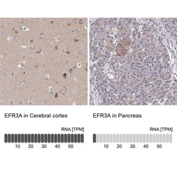
human ... EFR3A(23167)

Gene Information

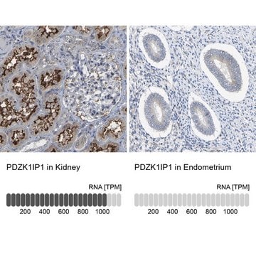
human ... PDZK1IP1(10158)

clone

polyclonal

clone

polyclonal

clone

polyclonal

clone

polyclonal

General description

The gene MYOF (myoferlin) encodes a type II membrane protein that shares homology with dysferlin. It is predominantly expressed in developing muscle and extensively in cardiac muscle. It is a member of the ferlin family of proteins. These proteins contain C2 domains that are important in calcium-mediated membrane fusion events. Myoferlin is found to be associated with both plasma membrane and nuclear membrane.

Immunogen

Myoferlin recombinant protein epitope signature tag (PrEST)

Application

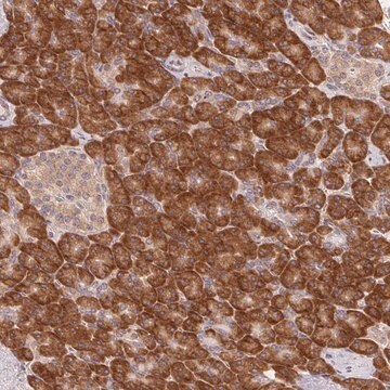
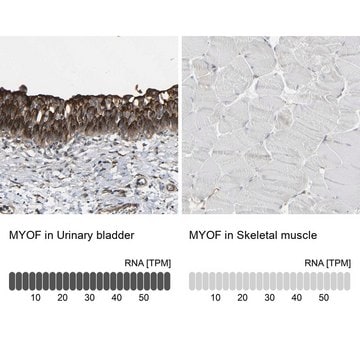
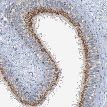
Anti-MYOF antibody produced in rabbit, a Prestige Antibody, is developed and validated by the Human Protein Atlas (HPA) project . Each antibody is tested by immunohistochemistry against hundreds of normal and disease tissues. These images can be viewed on the Human Protein Atlas (HPA) site by clicking on the Image Gallery link. The antibodies are also tested using immunofluorescence and western blotting. To view these protocols and other useful information about Prestige Antibodies and the HPA, visit sigma.com/prestige.

Biochem/physiol Actions

Myoferlin is involved in the fusion of endomembrane with the plasma membrane in muscle cells and endothelial cells. It forms a complex with dynamin-2 (Dyn-2) and promotes the surface expression of vascular endothelial growth factor receptor 2. The protein complex of myoferlin-Dyn-2-Cav-1 (caveolin-1) participates in the processes of membrane fusion and fission. Myoferlin is also implicated in receptor-dependent endocytosis. It is crucial for muscle development and regeneration, and its expression is found to increase in skeletal muscle on injury. It participates in vesicle recycling and its deficiency can lead to accumulation of transferrin in the perinuclear endocytic recycling compartment in the myoblasts. It regulates postnatal muscle growth mediated by IGF1 (insulin-like growth factor 1).

Features and Benefits

Prestige Antibodies® are highly characterized and extensively validated antibodies with the added benefit of all available characterization data for each target being accessible via the Human Protein Atlas portal linked just below the product name at the top of this page. The uniqueness and low cross-reactivity of the Prestige Antibodies® to other proteins are due to a thorough selection of antigen regions, affinity purification, and stringent selection. Prestige antigen controls are available for every corresponding Prestige Antibody and can be found in the linkage section.

Every Prestige Antibody is tested in the following ways:
  • IHC tissue array of 44 normal human tissues and 20 of the most common cancer type tissues.
  • Protein array of 364 human recombinant protein fragments.

Linkage

Corresponding Antigen APREST71990

Physical form

Solution in phosphate-buffered saline, pH 7.2, containing 40% glycerol and 0.02% sodium azide

Legal Information

Prestige Antibodies is a registered trademark of Merck KGaA, Darmstadt, Germany

Disclaimer

Unless otherwise stated in our catalog or other company documentation accompanying the product(s), our products are intended for research use only and are not to be used for any other purpose, which includes but is not limited to, unauthorized commercial uses, in vitro diagnostic uses, ex vivo or in vivo therapeutic uses or any type of consumption or application to humans or animals.

Not finding the right product?  

Try our Product Selector Tool.

Storage Class Code

10 - Combustible liquids

WGK

WGK 1

Flash Point(F)

Not applicable

Flash Point(C)

Not applicable

Personal Protective Equipment

dust mask type N95 (US), Eyeshields, Gloves

Certificates of Analysis (COA)

Search for Certificates of Analysis (COA) by entering the products Lot/Batch Number. Lot and Batch Numbers can be found on a product’s label following the words ‘Lot’ or ‘Batch’.

Need A Sample COA?

This is a sample Certificate of Analysis (COA) and may not represent a recently manufactured lot of this specific product.

Already Own This Product?

Find documentation for the products that you have recently purchased in the Document Library.

Visit the Document Library

Leonithas I Volakis et al.
PloS one, 9(2), e86110-e86110 (2014-03-04)
Cell migration plays a central role in the invasion and metastasis of tumors. As cells leave the primary tumor, they undergo an epithelial to mesenchymal transition (EMT) and migrate as single cells. Epithelial tumor cells may also migrate in a
Hailong Shi et al.
Frontiers in oncology, 12, 905230-905230 (2022-09-24)
In contrast to normal cells, in which reactive oxygen species (ROS) are maintained in redox equilibrium, cancer cells are characterized by ectopic ROS accumulation. Myoferlin, a newly identified oncogene, has been associated with tumor metastasis, intracellular ROS production, and energy
Kenji Harada et al.
Gastric cancer : official journal of the International Gastric Cancer Association and the Japanese Gastric Cancer Association, 24(6), 1264-1277 (2021-07-18)
The attainment of drug resistance in gastric cancer (GC) is a problematic issue. Although many studies have shown that cancer stem cells (CSCs) play an important role in the acquisition of drug resistance, there is no clinically available biomarker for
Arnaud Blomme et al.
Oncotarget, 7(50), 83669-83683 (2016-11-16)
Exosomes are communication mediators participating in the intercellular exchange of proteins, metabolites and nucleic acids. Recent studies have demonstrated that exosomes are characterized by a unique proteomic composition that is distinct from the cellular one. The mechanisms responsible for determining
Sandy Anania et al.
Cell communication and signaling : CCS, 22(1), 133-133 (2024-02-18)
Pancreatic ductal adenocarcinoma is an aggressive cancer type with one of the lowest survival rates due to late diagnosis and the absence of effective treatments. A better understanding of PDAC biology will help researchers to discover the Achilles' heel of

Our team of scientists has experience in all areas of research including Life Science, Material Science, Chemical Synthesis, Chromatography, Analytical and many others.

Contact Technical Service