Skip to Content
Merck
All Photos(5)

Key Documents

C2562

Sigma-Aldrich

Monoclonal Anti-Cytokeratin, pan (Mixture) antibody produced in mouse

clone C-11+PCK-26+CY-90+KS-1A3+M20+A53-B/A2, ascites fluid

Synonym(s):

Monoclonal Anti-Cytokeratin, pan (mixture), Panck Antibody, Panck Antibody - Monoclonal Anti-Cytokeratin, pan (Mixture) antibody produced in mouse

Sign Into View Organizational & Contract Pricing


About This Item

MDL number:
UNSPSC Code:
12352203
NACRES:
NA.41

biological source

mouse

Quality Level

conjugate

unconjugated

antibody form

ascites fluid

antibody product type

primary antibodies

clone

C-11+PCK-26+CY-90+KS-1A3+M20+A53-B/A2, monoclonal

contains

7% horse serum and 15 mM sodium azide as preservative

species reactivity

wide range

technique(s)

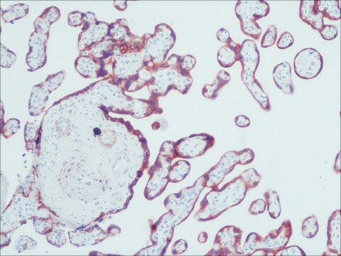
immunohistochemistry (formalin-fixed, paraffin-embedded sections): suitable using protease-digested sections of human or animal tissues
immunohistochemistry (frozen sections): suitable
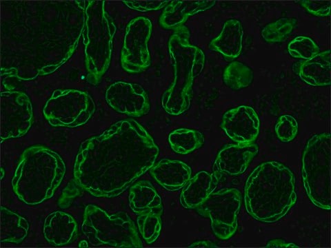
indirect immunofluorescence: 1:100 using protease-digested, formalin-fixed, paraffin-embedded sections of human or animal tissues
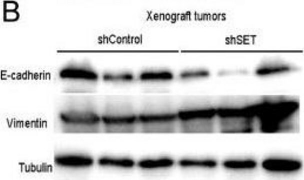
western blot: suitable

isotype

IgG1/IgG2a

shipped in

dry ice

storage temp.

−20°C

target post-translational modification

unmodified

Looking for similar products? Visit Product Comparison Guide

General description

Monoclonal Anti-Cytokeratin, pan (mouse IgG1 and IgG2a isotypes) is a mixture of monoclonal antibodies from the following clones: C-11, PCK-26, CY-90, KS1A3, M20, and A53-B/A2. Cytokeratins, a group comprising at least 29 different proteins, are characteristic of epithelial and trichocytic cells. Cytokeratins 1 (68 kDa), 4 (59 kDa), 5 (58 kDa), 8 (52 kDa), and 6 (56 kDa) are members of the type II neutral-to-basic subfamily expressed in cornified epithelia, non-cornified stratified squamous epithelia, stratified epithelia, simple epithelia and in tissues with natural or pathological turnover respectively. Cytokeratins 10 (56 kDa), 13 (54 kDa, and 18 (45 kDa) are members of the type I acidic subfamily expressed in cornified epithelia, non-cornified stratified squamous epithelia and simple epithelial cells respectively. Cytokeratin peptide 19 (40 kDa) is a type I keratin which can be expressed in both simple and in broad cells of stratifying epithelia at specific sites.

Specificity

This is a mixture of monoclonal anti-cytokeratin antibodies. It recognizes human cytokeratins 1,4,5,6,8,10,13,18, and 19. It is a broad spectrum reagent, which reacts specifically with a wide variety of normal, reactive, and neoplastic epithelial tissues. The antibody mixture reacts with simple, cornifying and non-cornifying squamous epithelia and pseudostratified epithelia. It does not react with non-epithelial normal human tissues. Increased staining intensity is seen following proteolytic treatment (protease unmasking).

Immunogen

mixture of several monoclonal cytokeratin clones.

Application

Monoclonal Anti-Cytokeratin, pan (Mixture) antibody produced in mouse is suitable for the following applications:
  • Immunohistochemistry (formalin-fixed, paraffin-embedded sections) using protease-digested sections of human or animal tissues.
  • Immunohistochemistry (frozen sections).
  • Indirect immunofluorescence (at a working dilution of 1:100 using protease-digested, formalin-fixed, paraffin-embedded sections of human or animal tissues).
  • Immunocytochemical labeling (immunofluorescence ) of cells.
  • Western blotting.

Biochem/physiol Actions

Cytokeratins are proteins of keratin-containing intermediate filaments that provide mechanical support and other additional functions in the epithelial cells. It is found in the intracytoplasmic cytoskeleton of epithelial tissue. Epithelial tissue expresses cytokeratin subunits in a specific and stable pattern. Cytokeratins along with vimentin are involved in cell proliferation, migration and differentiation of preodontoblasts and preameloblasts. The intermediate-sized filaments are abundant in human endothelial cells and are mostly of vimentin type.

Disclaimer

Unless otherwise stated in our catalog or other company documentation accompanying the product(s), our products are intended for research use only and are not to be used for any other purpose, which includes but is not limited to, unauthorized commercial uses, in vitro diagnostic uses, ex vivo or in vivo therapeutic uses or any type of consumption or application to humans or animals.

Not finding the right product?  

Try our Product Selector Tool.

Storage Class Code

10 - Combustible liquids

WGK

WGK 3


Certificates of Analysis (COA)

Search for Certificates of Analysis (COA) by entering the products Lot/Batch Number. Lot and Batch Numbers can be found on a product’s label following the words ‘Lot’ or ‘Batch’.

Already Own This Product?

Find documentation for the products that you have recently purchased in the Document Library.

Visit the Document Library

J M Pérez-Pomares et al.
Developmental dynamics : an official publication of the American Association of Anatomists, 210(2), 96-105 (1997-10-23)
A study about the hypothetical contribution of the epicardial cells to the subepicardial mesenchyme was carried out in Syrian hamster embryos of 9-12 days post coitum (dpc) and chick embryos of 3-5 days of incubation. In the epicardium and subepicardium
Qian Wang et al.
Journal of virology, 78(2), 821-833 (2003-12-25)
High-risk human papillomaviruses, such as human papillomavirus type 16 (HPV16), are the primary cause of cervical cancer. The HPV16 E1=E4 protein associates with keratin intermediate filaments and causes network collapse when expressed in epithelial cells in vitro. Here, we show
R Muñoz-Chápuli et al.
Differentiation; research in biological diversity, 64(3), 133-141 (1999-05-11)
The existence of the hemangioblast, a common progenitor of the endothelial and hematopoietic cell lineages, was proposed at the beginning of the century. Although recent findings seem to confirm its existence, it is still unknown when and how the hemangioblasts
Emmanuel Letavernier et al.
Nephrology, dialysis, transplantation : official publication of the European Dialysis and Transplant Association - European Renal Association, 24(2), 630-638 (2008-10-18)
The specific mTor inhibitor sirolimus has been implicated in the pathogenesis of renal glomerular lesions and nephrotic syndrome appearance after transplantation. Podocyte injury and focal segmental glomerulosclerosis have been related to sirolimus therapy in some patients but the pathways underlying
Structure and functions of keratin proteins in simple, stratified, keratinized and cornified epithelia
Bragulla HH and Homberger DG
Journal of Anatomy, 214(4), 516-559 (2009)

Our team of scientists has experience in all areas of research including Life Science, Material Science, Chemical Synthesis, Chromatography, Analytical and many others.

Contact Technical Service