Skip to Content
Merck
All Photos(3)

Key Documents

MABE1120

Sigma-Aldrich

Anti- HSPA8/Hsc70 Antibody, clone N69

clone N69, from mouse

Synonym(s):

HSPA8, Heat shock cognate 71 kDa protein, Heat shock 70 kDa protein 8, HSC70, HSC71, HSC73

Sign Into View Organizational & Contract Pricing


About This Item

UNSPSC Code:
12352203
eCl@ss:
32160702
NACRES:
NA.41

biological source

mouse

Quality Level

antibody form

purified immunoglobulin

antibody product type

primary antibodies

clone

N69, monoclonal

species reactivity

mouse, rat, bovine, human

technique(s)

flow cytometry: suitable
immunocytochemistry: suitable
immunohistochemistry: suitable
western blot: suitable

isotype

IgG1κ

NCBI accession no.

UniProt accession no.

shipped in

wet ice

target post-translational modification

unmodified

Gene Information

human ... HSPA8(3312)

Related Categories

General description

Heat shock cognate 71 kDa protein (UniProt P19120; also known as Heat shock 70 kDa protein 8, HSC70, HSC71, HSC73) is encoded by the HSPA8 (also known as HSC70, HSC73) gene (Gene ID 281831) in bovine species. The mammalian HSP70 family consists of the mitochondrial HSP75, the endoplasmic HSP78, the cytoplasmic HSC70 (a.k.a. HSC73 and HSP73) and its inducible counterpart HSP72 (a.k.a. HSP70). The latter two have similar structures and properties, and both of them bind misfolded, newly synthesized polypeptides in an ATP-dependent manner, reflecting their chaperonic activity. All cells constitutively express HSC70. On the other hand, the expression of HSP72 in normal cells is mainly stress inducible, although constitutively high HSP72 expressions are observed in malignant human tumors of various origins. The induced HSP72 expression enables stressed cells to better cope with the elevation of unfolded/denatured proteins and prevent apoptosis under various stress conditions.

Specificity

Clone N69 reacts with the constitutively expressed HSPA8/Hsc70 (HSC73), but not the heat schock-inducible HSP72 (Demidov, O.N., et al. (1999). Eur. J. Cardiothorac. Surg. 16(4):444-449).

Immunogen

Co-purified Hsc73 and Hsp72 proteins from bovine skeletal muscle.

Application

Anti- HSPA8/Hsc70 Antibody, clone N69 is an antibody against HSPA8/Hsc70 for use in Western Blotting, Immunocytochemistry, Flow Cytometry, Immunohistochemistry.
Research Category
Epigenetics & Nuclear Function
Research Sub Category
Nuclear Receptors
Western Blotting Analysis: 2.0 µg/mL of this antibody detected HSPA8/Hsc70 in 10 µg rat C6 glioma, human T47D ductal breast epithelial tumor, and human K-562 erythroleukemia cell lysates.
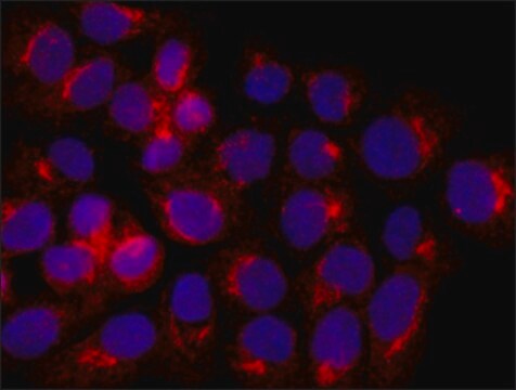
Immunocytochemistry Analysis: 2.0 µg/mL of a representative lot detected the constitutive HSPA8/Hsc70 expression in C6 rat glioma, B16 mouse melanoma, and K-562 human erythroblastoma cells (Courtesy of Dr. Irina V. Guzhova and Dr. Boris Margulis, Russian Academy of Sciences).
Flow Cytometry Analysis: 2.0 µg/mL of a representative lot detected the constitutive HSPA8/Hsc70 expression in C6 rat glioma, B16 mouse melanoma, and K-562 human erythroblastoma cells (Courtesy of Dr. Irina V. Guzhova and Dr. Boris Margulis, Russian Academy of Sciences).
Immunohistochemistry Analysis: A representative lot detected bovine HSPA8/Hsc70 in anterior hypothalamus frozen sections from rats received purified bovine Hsc70 and Hsp72 proteins via intracerebroventricular (i.c.v.) microinjections (Ekimova, I.V., et al. (2010). J. Neurochem. 115(4):1035-1044).
Western Blotting Analysis: A representative lot detected HSPA8/Hsc70 expression in MCF7 cells (Hoyer-Hansen, M., et al. (2005). Cell Death Differ. 12(10):1297-1309).
Western Blotting Analysis: A representative lot detected HSPA8/Hsc70 expression in human breast cancer cell lines (MDA-MB-468, MCF-7, BT-549, and SK-BR-3), non-tumorigenic breast-derived HBL-100 epithelial cells, and WI-38 normal lung fibroblasts (Nylandsted, J., et al. (2000). Proc. Natl. Acad. Sci. U. S. A. 97(14):7871-7876).
Western Blotting Analysis: A representative lot detected HSPA8/Hsc70 (HSC73) purified from bovine skeletal muscle and human myocardium, but not the co-purified HSP72 (Demidov, O.N., et al. (1999). Eur. J. Cardiothorac. Surg. 16(4):444-449).

Quality

Evaluated by Western Blotting in MCF7 cell lysate.

Western Blotting Analysis: 2.0 µg/mL of this antibody detected HSPA8/Hsc70 in 10 µg MCF7 cell lysate.

Target description

~71 kDa observed

Physical form

Format: Purified
Protein G Purified
Purified mouse monoclonal IgG1κ antibody in buffer containing 0.1 M Tris-Glycine (pH 7.4), 150 mM NaCl with 0.05% sodium azide.

Storage and Stability

Stable for 1 year at 2-8°C from date of receipt.

Other Notes

Concentration: Please refer to lot specific datasheet.

Disclaimer

Unless otherwise stated in our catalog or other company documentation accompanying the product(s), our products are intended for research use only and are not to be used for any other purpose, which includes but is not limited to, unauthorized commercial uses, in vitro diagnostic uses, ex vivo or in vivo therapeutic uses or any type of consumption or application to humans or animals.

Not finding the right product?  

Try our Product Selector Tool.

Storage Class Code

12 - Non Combustible Liquids

WGK

WGK 1

Flash Point(F)

Not applicable

Flash Point(C)

Not applicable


Certificates of Analysis (COA)

Search for Certificates of Analysis (COA) by entering the products Lot/Batch Number. Lot and Batch Numbers can be found on a product’s label following the words ‘Lot’ or ‘Batch’.

Already Own This Product?

Find documentation for the products that you have recently purchased in the Document Library.

Visit the Document Library

Masaharu Somiya et al.
Journal of extracellular vesicles, 7(1), 1440132-1440132 (2018-03-08)
Extracellular vesicles (EVs) deliver biologically active cargos from donor cells to recipient cells for intercellular communication. Since the existence of RNA cargo was discovered, EVs have been considered to be useful drug-delivery systems. Specifically, EVs from bovine milk (mEV) are
Attila Simor et al.
Molecular and cellular neurosciences, 79, 64-80 (2017-01-15)
Acute total sleep deprivation (SD) impairs memory consolidation, attention, working memory and perception. Structural, electrophysiological and molecular experimental approaches provided evidences for the involvement of sleep in synaptic functions. Despite the wide scientific interest on the effects of sleep on
Yan Zhou et al.
Glia, 72(4), 692-707 (2024-01-09)
Schwann cells (SCs), the primary glial cells of the peripheral nervous system, which have been identified in many solid tumors, play an important role in cancer development and progression by shaping the tumor immunoenvironment and supporting the development of metastases.
Shenée C Martin et al.
Pharmaceuticals (Basel, Switzerland), 17(3) (2024-03-28)
Sleep disruption is an expected component of aging and neurodegenerative conditions, including Alzheimer's disease (AD). Sleep disruption has been demonstrated as a driver of AD pathology and cognitive decline. Therefore, treatments designed to maintain sleep may be effective in slowing
Laura Le Gall et al.
Skeletal muscle, 10(1), 20-20 (2020-07-10)
Skeletal muscle is increasingly considered an endocrine organ secreting myokines and extracellular vesicles (exosomes and microvesicles), which can affect physiological changes with an impact on different pathological conditions, including regenerative processes, aging, and myopathies. Primary human myoblasts are an essential

Our team of scientists has experience in all areas of research including Life Science, Material Science, Chemical Synthesis, Chromatography, Analytical and many others.

Contact Technical Service